Category:Bones of the human foot
Zur Navigation springen
Zur Suche springen
| Medium hochladen | |||||
| Unterklasse von |
| ||||
|---|---|---|---|---|---|
| Ist Teil von |
| ||||
| |||||
- See also: Category:Tarsus, Category:Metatarsus, Category:Ankle
Unterkategorien
Es werden 14 von insgesamt 14 Unterkategorien in dieser Kategorie angezeigt:
In Klammern die Anzahl der enthaltenen Kategorien (K), Seiten (S), Dateien (D)
3
- 3D data of human foot bones (1 D)
H
M
- Metatarsal accessory bones (16 D)
O
P
- Photographs of human foot bones (12 D)
T
V
- Videos of human foot bones (3 D)
Medien in der Kategorie „Bones of the human foot“
Folgende 95 Dateien sind in dieser Kategorie, von 95 insgesamt.
-
 "Anatomia per uso...del disegno...", B. Genga, 1691 Wellcome L0009686.jpg 1.080 × 1.790; 634 KB
-
 202110 Dorsal view of bones of right foot.svg 512 × 512; 348 KB
-
 202110 Plantar view of bones of right foot.svg 512 × 512; 375 KB
-
 812 Bones of the Foot.jpg 1.119 × 817; 374 KB
-
 BodyParts3D Foot bones.stl 5.120 × 2.880; 2,28 MB
-
 Bones of the foot, ankle and knee joint; eight figures. Penc Wellcome V0008239.jpg 3.331 × 2.313; 3,57 MB
-
 Bones of the foot, forearm, and hand. Crayon manner print by Wellcome V0007883ER.jpg 1.719 × 1.180; 1,05 MB
-
 Bones of the human skeleton - ribcage and foot Wellcome V0008819.jpg 2.492 × 3.708; 4 MB
-
 Bound feet (X-ray).jpg 3.709 × 2.892; 4,11 MB
-
 Calcaneus Fracture.jpg 2.270 × 1.542; 256 KB
-
 Calcar Calcanei 01.jpg 2.270 × 1.604; 1,27 MB
-
 Calcar Calcanei 02.jpg 2.272 × 1.704; 1,75 MB
-
 ChineseLadieFoot.gif 1.837 × 952; 74 KB
-
 Cunningham’s Text-book of Anatomy (1914) - Fig 270.png 1.644 × 1.005; 1,32 MB
-
 Cunningham’s Text-book of Anatomy (1914) - Fig 272.png 666 × 1.388; 1,51 MB
-
 Cunningham’s Text-book of Anatomy (1914) - Fig 277.png 1.242 × 632; 492 KB
-
 Dixon's Manual of human osteology (1912) - Fig 083.png 1.371 × 2.001; 758 KB
-
 Dixon's Manual of human osteology (1912) - Fig 088.png 1.308 × 2.064; 1,19 MB
-
 Dixon's Manual of human osteology (1912) - Fig 098.png 2.196 × 768; 969 KB
-
 Dixon's Manual of human osteology (1912) - Fig 099.png 1.530 × 1.959; 940 KB
-
 Dixon's Manual of human osteology (1912) - Fig 100.png 1.362 × 2.082; 908 KB
-
 Em - Homo sapiens - 40.jpg 1.855 × 2.782; 678 KB
-
 Foot (10825992734).jpg 1.180 × 2.930; 431 KB
-
Foot Bone Anatomy by Jason Christian.webm 28 s, 1.280 × 720; 4,92 MB
-
 Foot bones - tarsus, metatarsus and phalanges.jpg 960 × 720; 99 KB
-
Foot bones overview.webm 2 min 27 s, 1.077 × 606; 12,55 MB
-
 Foot bones RO.png 935 × 661; 93 KB
-
Foot Bones Walk-thru by Bob Myers.webm 6 min 32 s, 640 × 480; 8,68 MB
-
 Foot bones.jpg 657 × 1.305; 85 KB
-
 Foot4 (10818502746).jpg 2.268 × 1.624; 510 KB
-
 Fulcrum into skeletal foot.zero balancing tutorial.JPG 3.888 × 2.592; 3,18 MB
-
 Fuss eines 17 jährigen Jünglings MET DP262475.jpg 2.847 × 3.889; 1,88 MB
-
 Gerrish's Text-book of Anatomy (1902) - Fig. 198.png 1.436 × 2.672; 1,96 MB
-
 Gerrish's Text-book of Anatomy (1902) - Fig. 199.png 1.534 × 2.625; 684 KB
-
 Gerrish's Text-book of Anatomy (1902) - Fig. 200.png 1.540 × 2.624; 2,33 MB
-
 Gerrish's Text-book of Anatomy (1902) - Fig. 201.png 1.852 × 2.684; 971 KB
-
 Gerrish's Text-book of Anatomy (1902) - Fig. 202.png 1.952 × 724; 888 KB
-
 Gerrish's Text-book of Anatomy (1902) - Fig. 203.png 1.922 × 612; 744 KB
-
 Hand foot (10851018025).jpg 1.632 × 2.464; 744 KB
-
 HKMMS Bound Feet y60318.jpg 1.920 × 2.560; 1,63 MB
-
 Holden's human osteology (1899) - Plt36.png 2.772 × 1.136; 1,96 MB
-
 Holden's human osteology (1899) - Plt37.png 1.951 × 2.152; 3 MB
-
 Huesos del pie.jpg 400 × 375; 27 KB
-
 Imaginea 4.JPG 1.704 × 2.272; 1,82 MB
-
 Jacopo Berengarius da Carpi, Isagoge breves Wellcome L0031353.jpg 1.728 × 1.278; 1,07 MB
-
 Leonardo Anatomical Studies.jpg 485 × 600; 121 KB
-
 Leprosy deformedfeet.JPG 2.304 × 1.536; 368 KB
-
 Merkel's Human Anatomy (1913) - Vol 3 - Fig 127.png 1.113 × 1.476; 445 KB
-
 Merkel's Human Anatomy (1913) - Vol 3 - Fig 128.png 1.329 × 1.335; 994 KB
-
 Morris' human anatomy (1898) - Fig 166.png 1.396 × 2.304; 1,94 MB
-
 Morris' human anatomy (1898) - Fig 167.png 1.664 × 2.340; 2,31 MB
-
 Morris' human anatomy (1933) - Fig 276.png 1.924 × 2.717; 2,77 MB
-
 Morris' human anatomy (1933) - Fig 277.png 2.123 × 2.815; 3,39 MB
-
 Os cuboideum.jpeg 390 × 253; 49 KB
-
 Ospied-ar.jpg 418 × 366; 37 KB
-
 Ospied.jpg 418 × 366; 20 KB
-
 Ossa pedis (sin) - calcaneus (medial view).jpg 4.608 × 3.456; 4,53 MB
-
 Ossa pedis (sin) - lateral view.jpg 4.608 × 3.456; 4,61 MB
-
 Ossa pedis (sin) - medial view.jpg 4.608 × 3.456; 2,05 MB
-
 Ossa pedis (sin) - medialis et superior.jpg 4.608 × 4.424; 3,33 MB
-
 Ossa pedis (sin) - ossa metatarsi (medial view).jpg 4.608 × 3.456; 4,01 MB
-
 Ossa pedis (sin) - ossa metatarsi (superior view).jpg 4.608 × 3.456; 4,61 MB
-
 Ossa pedis (sin) - superior view 2.jpg 4.608 × 3.456; 2,25 MB
-
 Ossa pedis (sin) - superior view.jpg 4.608 × 3.456; 3,11 MB
-
 PersonalHygeine p122.jpg 2.302 × 3.325; 1,47 MB
-
 PersonalHygeine p123.jpg 3.600 × 1.398; 1,21 MB
-
 Pie - 2.png 603 × 366; 22 KB
-
 RadZepMov CT 3D human Foot Bone SHR.jpeg 400 × 400; 29 KB
-
 Richer - Anatomie artistique, 2 p. 36.png 6.744 × 8.925; 4,86 MB
-
 Sesamoidbone.png 1.376 × 880; 409 KB
-
 SesamoidBonesOfFoot.svg 1.362 × 1.099; 388 KB
-
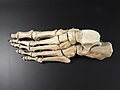
 Skeleton foot.JPG 2.288 × 1.712; 2,25 MB
-
 Skeleton of a foot. Wellcome M0006912.jpg 4.110 × 2.586; 2,9 MB
-
 Sketches of anatomy Wellcome L0011853.jpg 1.270 × 1.578; 791 KB
-
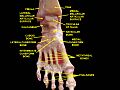
 Slide31DEN.JPG 960 × 720; 66 KB
-
 Slide5ecce - Navicular bone.png 960 × 720; 456 KB
-
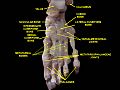
 Slide5ecce-ar.jpg 960 × 720; 132 KB
-
 Slide5ecce.JPG 960 × 720; 71 KB
-
 Slide6CEC5 - Navicular bone.png 960 × 720; 781 KB
-
 Slide6CEC5.JPG 960 × 720; 87 KB
-
 Sobo 1909 225.png 1.470 × 2.307; 9,72 MB
-
 Sobo 1909 227.png 1.884 × 1.413; 7,63 MB
-
 Sobo 1909 228.png 2.799 × 1.590; 12,75 MB
-
 Spalteholz's Hand-Atlas of Human Anatomy (1906) - Vol 1 - Fig 202.png 2.336 × 3.192; 1,42 MB
-
 Spalteholz's Hand-Atlas of Human Anatomy (1906) - Vol 1 - Fig 203.png 2.368 × 3.208; 2,11 MB
-
 Tape9.png 316 × 250; 134 KB
-
 Tarsalia accessoria.png 400 × 400; 15 KB
-
 Tarsus.png 400 × 400; 14 KB
-
 Tarsus2.png 400 × 400; 17 KB
-
 Testut's Treatise on Human Anatomy (1911) - Vol 1 - Fig 401.png 984 × 2.388; 925 KB
-
 Testut's Treatise on Human Anatomy (1911) - Vol 1 - Fig 403.png 1.064 × 2.364; 1,39 MB
-
 True bound foot of a chinese woman 43 years Wellcome V0031188.jpg 2.350 × 3.078; 2,69 MB
-
 Bones of the foot; six figures. Pencil drawing, ca. 1811. Wellcome V0008240.jpg 3.500 × 2.046; 2,97 MB
-
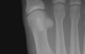
 X-ray.jpg 1.036 × 775; 87 KB
-
 НЭС. Обувь 1.jpg 105 × 216; 9 KB


.jpg)


_-_Fig_270.png)
_-_Fig_272.png)
_-_Fig_277.png)
_-_Fig_083.png)
_-_Fig_088.png)
_-_Fig_099.png)
_-_Fig_100.png)


.jpg)


_-_Fig._198.png)
_-_Fig._199.png)
_-_Fig._200.png)
_-_Fig._201.png)
.jpg)

_-_Plt37.png)


_-_Vol_3_-_Fig_127.png)
_-_Vol_3_-_Fig_128.png)
_-_Fig_166.png)
_-_Fig_167.png)
_-_Fig_276.png)
_-_Fig_277.png)


_-_calcaneus_(medial_view).jpg)
_-_lateral_view.jpg)
_-_medial_view.jpg)
_-_medialis_et_superior.jpg)
_-_ossa_metatarsi_(medial_view).jpg)
_-_ossa_metatarsi_(superior_view).jpg)
_-_superior_view_2.jpg)
_-_superior_view.jpg)


_-_Vol_1_-_Fig_202.png)
_-_Vol_1_-_Fig_203.png)


_-_Vol_1_-_Fig_403.png)


_-_Fig_098.png)
.jpg)
_-_Fig._202.png)
_-_Fig._203.png)
_-_Plt36.png)
_-_Vol_1_-_Fig_401.png)