Category:Cytokines
Vai alla navigazione
Vai alla ricerca
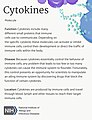
vasta e ampia categoria di piccole proteine (~5-20 kDa) che sono importanti nella segnalazione cellulare | |||||
| Carica un file multimediale | |||||
| Istanza di |
| ||||
|---|---|---|---|---|---|
| Sottoclasse di |
| ||||
| Distinto da | |||||
| |||||
English: Cytokines; Interferon, Interleukin, Growth factor, Tumor necrosis factor, etc.
日本語: サイトカイン;インターフェロン、インターロイキン、成長因子、腫瘍壊死因子など
Sottocategorie
Questa categoria contiene le 13 sottocategorie indicate di seguito, su un totale di 13.
Pagine nella categoria "Cytokines"
Questa categoria contiene un'unica pagina, indicata di seguito.
File nella categoria "Cytokines"
Questa categoria contiene 75 file, indicati di seguito, su un totale di 75.
-
De-Zytokin.ogg 2,0 s; 19 KB
-
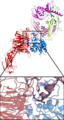
 1b53.png 428 × 335; 70 KB
-
 1ESR Human Monocyte Chemotactic Protein-2 01.png 2 400 × 1 673; 769 KB
-
 1ESR Human Monocyte Chemotactic Protein-2 02.png 2 400 × 1 673; 349 KB
-
 1ESR Human Monocyte Chemotactic Protein-2.png 1 178 × 850; 111 KB
-
 1ESR.pdb1.png 1 436 × 759; 219 KB
-
 1hum.png 359 × 241; 49 KB
-
 1JE4 Monomeric Variant Of The Chemokine Mip-1beta01.png 2 400 × 1 673; 865 KB
-
 1JE4 Monomeric Variant Of The Chemokine Mip-1beta02.png 2 400 × 1 673; 296 KB
-
 1JE4 Monomeric Variant Of The Chemokine Mip-1beta03.png 2 400 × 1 673; 1 MB
-
 1JE4 Monomeric Variant Of The Chemokine Mip-1beta04.png 2 400 × 1 673; 1,16 MB
-
 1JE4 Monomeric Variant Of The Chemokine Mip-1beta05.png 2 400 × 1 673; 919 KB
-
 1JE4Monomeric Variant Of The Chemokine Mip-1beta.png 1 242 × 682; 99 KB
-
 2KLL.pdb.png 1 436 × 759; 249 KB
-
 3JVF.pdb.jpg 492 × 489; 45 KB
-
 3KKH.pdb.png 837 × 632; 160 KB
-
 3LTQ.pdb.png 1 436 × 759; 173 KB
-
 4JVF.pdb.jpg 492 × 489; 36 KB
-
 5JVF.pdb.jpg 492 × 489; 38 KB
-

-
 AVb6-TGFb1 binding.png 1 075 × 2 013; 1,46 MB
-
 BDNF 1BND.jpg 2 970 × 1 017; 419 KB
-
 Biochem Mol..png 252 × 168; 17 KB
-
 Chtx-Deu8.png 960 × 720; 58 KB
-
 Chtx-Deu9.png 960 × 720; 81 KB
-
 Chtx-wikifr-9.png 960 × 720; 80 KB
-
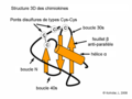
 ChtxChemkinStr2.jpg 1 084 × 889; 84 KB
-
 ChtxChemkinStr2.png 960 × 653; 25 KB
-
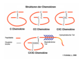
 ChtxChemokineStruct Dutchtxt.png 720 × 540; 6 KB
-
 ChtxChemokineStruct.png 720 × 500; 7 KB
-
 ChtxChimiokineStruct.png 1 200 × 900; 84 KB
-
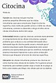
 Citochine.png 344 × 292; 37 KB
-
 Citocina (Cytokine) (35795300774).jpg 1 149 × 1 681; 269 KB
-
 Cytokine profile in Sjogren’s syndrome (SS).png 625 × 452; 185 KB
-
 Cytokine release following SARS-Cov-2 infection resulting in ARDS related to COVID-19.png 4 310 × 3 357; 1,88 MB
-
 Cytokine release.jpg 1 920 × 1 080; 1,46 MB
-
 Cytokines (34681563363).jpg 1 149 × 1 497; 216 KB
-
 Edema Hands 01.jpg 1 013 × 646; 287 KB
-
 Esquema de les interaccions establertes per LIGHT i consequencies.jpg 1 152 × 720; 89 KB
-
 Esquema degranulacion celula cebada.jpg 654 × 490; 60 KB
-
 Esquema que mostra les consecuències de la unió de DcR3 a LIGHT.png 598 × 319; 32 KB
-
 Exercise induced Skeletal muscle secretome.jpg 1 950 × 1 986; 367 KB
-
 Familias de receptores de citocinas.jpg 1 454 × 1 080; 178 KB
-
 Fcell-08-00677-g001.jpg 2 474 × 1 378; 377 KB
-
 Fimmu-11-01648-g001.jpg 1 084 × 943; 337 KB
-
 Fimmu-11-01648-g002.jpg 957 × 869; 324 KB
-
 Fimmu-11-01648-g003.jpg 957 × 680; 171 KB
-
 Fimmu-11-01648-g004.jpg 953 × 639; 190 KB
-
 Fluorospot staining.png 978 × 326; 237 KB
-
 Fluorospotassay.png 700 × 319; 43 KB
-
 Fluorospotprincipfinal.jpg 1 243 × 801; 214 KB
-
 Funciones de las citocinas.png 2 480 × 3 508; 976 KB
-
 GMCSF Crystal Structure.rsh.png 900 × 750; 236 KB
-
 IL13 Solution Structure.rsh.png 900 × 750; 201 KB
-
 IL17F 1JPY.png 960 × 720; 150 KB
-
 IL19 Crystal Structure.png 756 × 753; 205 KB
-
 IL2 Crystal Structure.png 900 × 750; 204 KB
-
 IL5 Crystal Structure.rsh.png 900 × 750; 248 KB
-
 Inflammatoire reflex.svg 500 × 500; 219 KB
-
 Interleukin 16.png 773 × 693; 63 KB
-
 Interleukin-21-2OQP.png 1 533 × 801; 285 KB
-
 Leg Edema 01.jpg 591 × 403; 105 KB
-
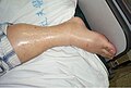 Leg Edema 02.jpg 599 × 403; 99 KB
-
 Manifestations cliniques et biologiques du syndrome de libération des cytokines.jpg 1 423 × 1 520; 348 KB
-
 Mifamurtide synthesis.png 1 375 × 1 884; 58 KB
-
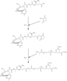 Mifamurtide synthesis2.png 1 552 × 1 891; 58 KB
-
 Muscle-Organ Crosstalk (Myokine).jpg 1 980 × 1 850; 348 KB
-
 Parasite160015-fig6 - Induction of proinflammatory cytokines in mice.png 945 × 1 439; 194 KB
-
 Penile Edema 01.jpg 597 × 413; 171 KB
-
 Penile Edema 02.jpg 602 × 412; 95 KB
-
 Physiopathologie et grade du syndrome de libération des cytokines.webp 1 944 × 1 408; 425 KB
-
 Receptores citoquinas IL-2.png 3 508 × 2 480; 986 KB
-
 Señalizacion citocinas.png 2 480 × 3 508; 900 KB
-
 Strong Hydrogen Bond between Asn34 and Arg82 of Different Monomers.png 2 936 × 1 722; 3,61 MB
-
 Structure and activation of cytokine.JPG 2 848 × 1 478; 1,42 MB


_(35795300774).jpg)
.png)


.jpg)


.jpg)


