Category:Livers
Gezinti kısmına atla
Arama kısmına atla

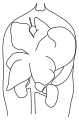
vertebrate organ involved in metabolism  Hígado de una oveja: 1. Lóbulo derecho, 2 Lóbulo izquierdo, 3 Lóbulo caudado, 4 Lóbulo cuadrado, 5 Arteria hepática y vena porta, 6 Nódulos linfáticos, 7 Vesícula biliar. | |||||
| Medya yükle | |||||
| Nedir |
| ||||
|---|---|---|---|---|---|
| Alt kümesidir |
| ||||
| Parçası | |||||
| Öncesinde |
| ||||
| Kısmen çakıştığı | |||||
| |||||
Alt kategoriler
Bu kategoride, aşağıdaki 13 alt kategori dahil toplam 13 alt kategori vardır.
-
C
- Couinaud classification of liver (18 F)
F
H
I
- Intrahepatic bile ducts (5 F)
L
- Liver of Piacenza (22 F)
P
"Livers" kategorisindeki ortam dosyaları
Bu kategoride yer alan toplam 77 dosyanın 77 adedi aşağıdadır.
-
 3D Medical Animation liver parts.jpg 1.920 × 1.080; 763 KB
-
 Alimentary Canal in a human embryo of the 3rd week.jpg 967 × 624; 250 KB
-
 Anatomic and histopathological aspects of FT organs human.jpg 3.948 × 1.292; 4,99 MB
-
 Ballooning degeneration high mag cropped annotated.jpg 1.700 × 1.124; 1,16 MB
-
 Bile recycling.png 432 × 453; 61 KB
-
 Bile recycling.svg 399 × 424; 14 KB
-
-
 Bottle of 'Chologestin', United States, 1890-1930 Wellcome L0058557.jpg 2.832 × 4.256; 1,64 MB
-
 Branches of coeliac trunk and hepatic arteries on angiography.png 791 × 1.023; 660 KB
-
 C2orf72 Ortholog Space Wiki Image.png 1.667 × 1.109; 146 KB
-
 C2orf72 Orthologs List.png 807 × 616; 305 KB
-
 CT Scan Thorax Liver.jpg 917 × 895; 193 KB
-
 EB1911 - Liver - Fig. 1.—The Liver from below and behind.jpg 639 × 699; 257 KB
-

-

-

-

-
 ELPA logo.png 597 × 274; 66 KB
-
 Estructura quilomicrón.jpg 1.654 × 1.654; 210 KB
-
 F. Glisson, plate I,"Anatomia hepatis" Wellcome L0013985.jpg 1.480 × 1.256; 550 KB
-
 Free Radical Toxicity.svg 603 × 652; 107 KB
-
 GSH LIVER image.png 650 × 413; 359 KB
-

-
 Hepatocellular Carcinoma- A Practical Approach 1st Edition.jpg 350 × 499; 25 KB
-
 Hepatocyte Culture.tif 894 × 653; 1,47 MB
-
 Hepatomegaly - CT single angle.jpg 512 × 512; 31 KB
-
 Hilum of the liver-ar.png 745 × 513; 545 KB
-
 Histological appearance of the FT liver with normal PVS human.jpg 1.978 × 1.078; 2,72 MB
-
 Histopathological and ultrasound aspect of normal FT liver human.jpg 4.016 × 1.092; 1,95 MB
-
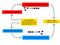 Homeostasis of blood sugar.png 1.200 × 900; 85 KB
-
 HPA RNA-Seq normal tissues C2Orf72 Expression Profile July 16 2022.png 2.241 × 840; 60 KB
-
 I-TASSER C2Orf72 Summer 2021 structure prediction.png 977 × 1.028; 473 KB
-
 Image of lungs and liver.jpg 4.000 × 1.800; 1,91 MB
-
 Intestinal carbohydrate digestion and absorption.jpg 3.405 × 1.350; 2,09 MB
-
 Kanzou.jpg 515 × 425; 74 KB
-

-
 Level of obstruction in Portal hypertension.svg 409 × 110; 18 KB
-
 Livartil P-FG-ES-03290.jpg 3.485 × 5.290; 3,49 MB
-
 Liver 04 Couinaud classification.svg-ar.png 2.608 × 1.564; 1,37 MB
-
 Liver development and in vitro hepatic differentiation of embryonic stem cells.jpg 1.152 × 1.124; 514 KB
-
 Liver example.png 1.600 × 1.200; 132 KB
-
 Liver regeneration-ar.png 1.292 × 362; 72 KB
-
 Maintenance of blood glucose during fasting.jpg 2.821 × 1.300; 1,89 MB
-
 Mallory body high mag cropped annotated.jpg 1.118 × 1.216; 595 KB
-
 Marina explaining Science.jpg 3.000 × 4.000; 1,14 MB
-
 Metabolism of lipoproteins.jpg 3.418 × 2.131; 2,49 MB
-
-
-
-
-
-
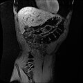 MRI of torso.jpg 512 × 512; 100 KB
-
 Museum Boğazkale 36.jpg 3.904 × 3.352; 9,39 MB
-
 Oddratio1figure3.pdf 2.133 × 1.641; 61 KB
-
 Oddratio2(Figure4).pdf 2.133 × 1.641; 224 KB
-
 Prometheus s3 V0041000 V0041860 full.jpg 3.220 × 2.336; 3,1 MB
-
 Reticular Cells.jpg 400 × 308; 46 KB
-
 S9 fraction.JPG 2.304 × 1.728; 2,31 MB
-
 Slide6CHA.JPG 960 × 720; 103 KB
-
 The American journal of anatomy (1914) (17532090404).jpg 2.304 × 3.238; 1,88 MB
-
 The American journal of anatomy (1914) (18155808001).jpg 2.172 × 3.048; 1,74 MB
-
 The liver, Chinese woodcut, Ming period Wellcome L0034722.jpg 2.005 × 3.277; 2,09 MB
-
 The liver.png 315 × 228; 6 KB
-
 Three anatomical figures from Tibet Wellcome V0036134.jpg 2.691 × 3.215; 3,36 MB
-
 Three Graces, Liverpool.jpg 4.624 × 3.468; 4,49 MB
-
 Tianxingju.JPG 1.198 × 786; 405 KB
-
 Ultrasound liver right lobe and right kidney.jpg 960 × 720; 237 KB
-
 Von Meyenburg complex cropped.tif 554 × 403; 166 KB
-
 Vuitton et al - International consensus on terminology - parasite200043-fig3.png 1.594 × 1.853; 731 KB
-
 Wiki gráfica hígado.png 752 × 721; 75 KB
-
 XMARS.JPG 2.736 × 3.648; 3,14 MB
-
 Подтверждение собственности фотографии по препарату( Печень).jpeg 2.448 × 3.264; 1,27 MB
-
 Правая и левая треугольная связки печени у кошки.jpg 5.152 × 3.864; 7,7 MB
-
 انواع مویرگ ها در جانوران.jpg 1.960 × 739; 217 KB
-
 سیاهرگ های کبد.jpg 584 × 396; 112 KB
-
 کالبد شناسی میکروسکوپی کبد.jpg 721 × 824; 198 KB
-
 کالبدشناسی میکروسکوپی کبد.jpg 721 × 824; 193 KB


.jpg)


_(17532090404).jpg)
_(18155808001).jpg)


.jpeg)


