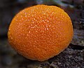
Category:Protista
Перайсьці да навігацыі
Перайсьці да пошуку
каралеўства эўкарыётных арганізмаў, якія не адносяцца да жывёлаў, расьлінаў і грыбоў   | |||||
| Загрузіць мэдыя | |||||
| Гук вымаўленьня | |||||
|---|---|---|---|---|---|
| Асобны выпадак панятку |
| ||||
| Падкляса ад | |||||
| Дата заснаваньня / стварэньня |
| ||||
| |||||
| |||||
| Міжнародная навуковая назва |
| ||||
| |||||
Падкатэгорыі
Гэтая катэгорыя зьмяшчае наступныя 22 падкатэгорыі з 22 агулам.
Старонкі ў катэгорыі «Protista»
Гэтая катэгорыя ўтрымлівае толькі адну старонку.
Файлы ў катэгорыі «Protista»
Паказаныя 200 файлаў гэтай катэгорыі з 349.
(папярэдняя старонка) (наступная старонка)-
 3D-fluorescence imaging for high throughput analysis of microbial eukaryotes.jpg 5098 × 3285; 1,39 Мб
-
 41467 2023 40657 Fig1.webp 1750 × 1199; 358 кб
-
 709 2021 1665 Fig13 HTML.webp 1524 × 2195; 1,05 Мб
-
 709 2021 1665 Fig13p.jpg 252 × 464; 22 кб
-
 709 2021 1665 Fig13q.jpg 518 × 418; 32 кб
-
 709 2021 1665 Fig13r.jpg 347 × 367; 20 кб
-
 709 2021 1665 Fig13s.jpg 440 × 425; 28 кб
-
 709 2021 1665 Fig13t.jpg 256 × 201; 10 кб
-
 709 2021 1665 Fig13u.jpg 290 × 209; 11 кб
-
 709 2021 1665 Fig13v.jpg 211 × 174; 7 кб
-
 709 2021 1665 Fig13w.jpg 257 × 267; 13 кб
-
 Acanthostomella Jörgensen, 1927.png 333 × 397; 65 кб
-
 Acineta patula Claparede & Lachmann, 1881 in Clap. & Lach 1860.jpg 1251 × 1890; 297 кб
-

-
 Acinetopsis rara, Robin 1879.jpg 273 × 1040; 28 кб
-
 Acropisthium mutabile in Pritchard 1861.png 462 × 866; 174 кб
-
 Actinobolina in Kahl 1930.png 606 × 754; 411 кб
-
 Actinomonas mirabilis, flagellate.jpg 535 × 1000; 75 кб
-
 Alga Fucus vesiculosus 090510 37.jpg 1500 × 932; 513 кб
-

-
 Allantosoma Gassovsky, 1919.jpg 597 × 636; 72 кб
-
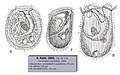
 Amphisiella in Kahl 1932.png 666 × 1506; 649 кб
-
 Ancyromonas@SciELO.jpg 1768 × 825; 90 кб
-
 Anoplophrya branchiarum Stein in Bütschli, 1887-1889.jpg 1096 × 1030; 365 кб
-
 Anoplophrya maupasi in Cépède 1910 Planche XIII.jpg 994 × 1384; 574 кб
-
 Anthophysa colonies.jpg 672 × 1000; 120 кб
-
 Atopochilodon arenifer in Kahl 1935.png 402 × 544; 187 кб
-

-
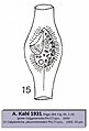
 Balanion comatum Wulff, 1919.png 872 × 1196; 588 кб
-
 Balladyna et Balladynopsis in Kahl 1932.png 1049 × 927; 617 кб
-
 Blepharisma in Kahl 1932.png 831 × 1328; 845 кб
-
 Blepharocorys bovis Dogiel, 1926.jpg 1094 × 1427; 198 кб
-
 Buetschliella (Bütschliella) opheliae Awerinzew, 1908.jpg 1444 × 2804; 626 кб
-
 Bursaridium Lauterborn, 1894 in Kahl 1932.jpg 1167 × 726; 232 кб
-
 Caenomorpha sp. in Kahl 1930.jpg 378 × 559; 18 кб
-
 Calyptotricha. pleuronemoides Phillips, 1882 in Kahl 1931.jpg 575 × 847; 68 кб
-
 Certesia quadrinucleata Fabre-Domergue, 1885.png 706 × 1404; 726 кб
-
 Chaetospira in Kahl 1932.png 926 × 1407; 521 кб
-
 Chattonidium setense in H.R. Pinkham 1953.png 747 × 729; 491 кб
-
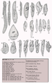
 Chilodonella (45 images) in Kahl 1931 et 1935.png 1920 × 1500; 1,92 Мб
-
 Chilodontopsis in Kahl 1931 et 1935.jpg 1138 × 1106; 280 кб
-
 Chlamydodon in Kahl 1931.png 1234 × 1263; 1,54 Мб
-
 Choanophrya infundibulifera Hartog, 1902.jpg 1116 × 1268; 258 кб
-
 Chromista structure.jpg 685 × 260; 105 кб
-
 Cicle vital de Cyclospora Cayetanensis.png 2730 × 3627; 2,26 Мб
-
 Ciliophrys DIC.jpg 1047 × 1000; 238 кб
-
 Cinetochilum margaritaceum (Ehrenberg, 1831) Perty, 1849.jpg 584 × 692; 74 кб
-
 Cinq espèces de Cycloposthium in A. Strelkow, 1928.jpg 1720 × 2940; 714 кб
-
 Cladogram for some unicellular eukaryotes.webp.jpg 1183 × 1500; 251 кб
-
 Cladotricha Gajewskaja, 1926 in Kahl 1932.jpg 333 × 593; 68 кб
-
 Clathrostoma Penard, 1922 in Kahl 1931.jpg 1233 × 916; 292 кб
-
 Clevelandella & Paraclevelandia in Richard Pinkham 1953.png 885 × 930; 640 кб
-
 Climacostomum virens in Bütschli & Conrad 1889.png 637 × 1073; 490 кб
-
 Coelosomides marina Anigstein 1912 in Kahl 1930.jpg 379 × 733; 65 кб
-
 Cohnilembus (Lembus) punctatus Kahl, 1933.jpg 614 × 972; 77 кб
-
 Collinia circulans (Balbiani, 1885) Cépède, 1910.jpg 754 × 854; 226 кб
-
 Cometodendron digitatum Swarczewsky.jpg 578 × 749; 77 кб
-
 Conchophthirus in Kahl 1931.png 917 × 1215; 994 кб
-
 Conchophthirus mytili Morgan, 1925.jpg 754 × 1130; 258 кб
-
 Conchophyllum caryoclada, (Kidder, 1933) Raabe, 1936.png 480 × 786; 427 кб
-
 Conidophrys pilisuctor Chatton & Lwoff, 1934.jpg 1194 × 1350; 530 кб
-
 Corynophrya divers schémas de 1878 à 1943 (classés par année).jpg 1437 × 1624; 338 кб
-
 Corynophrya divers schémas de 1878 à 1943.jpg 1437 × 1716; 342 кб
-
 Cryptochilum in Shevi︠akov 1896.jpg 1346 × 1406; 603 кб
-
 Cryptopharynx in Kahl 1931.jpg 512 × 752; 136 кб
-
 Ctedoctema acanthocrypta Stokes,1884 in Kahl 1931.jpg 409 × 855; 67 кб
-
 Cyathodinium conicum da Cunha, 1914.jpg 464 × 990; 88 кб
-
 Cyclogramma tricirrata.jpg 732 × 948; 128 кб
-
 Cycloposthium in Kudo 1946.jpg 1024 × 998; 256 кб
-
 Cyrtocaryum halosydnae Fauré-Fremiet & Mugard, 1949.jpg 1277 × 940; 368 кб
-
 Cyrtolophosis mucicola Stokes, 1885.png 939 × 733; 270 кб
-
 Dactylophrya collinii Gajewskaja, 1929.jpg 424 × 974; 110 кб
-
 Deformed Colopoda sp.png 2634 × 2004; 5,09 Мб
-
 Deltopylum rhabdoides d'après Mugard, 1946.jpg 1171 × 963; 245 кб
-
 Dendrocometes paradoxus Stein, 1852.jpg 1125 × 1600; 280 кб
-
 Dendrosoma radians Ehrenberg, 1838.jpg 1024 × 890; 239 кб
-
 Dendrosomides paguri Collin, 1906.jpg 881 × 664; 172 кб
-

-

-

-

-

-

-
-

-
 Die Radiolarien (Rhizopoda radiaria) - eine Monographie (1887) (20754175700).jpg 2232 × 2832; 1,81 Мб
-
-
 Discocephalus rotatorius Ehrenberg, 1829.png 508 × 592; 151 кб
-
 Discophrya elongata in Collin 1911.jpg 1140 × 1318; 299 кб
-
 Divers genres et espèces de Ditoxidae in R.R. Kudo 1931 & 1946.jpg 1518 × 2172; 562 кб
-
 Divers Mesnilella Cépède 1910.jpg 1776 × 2622; 638 кб
-
 Diverses espèces de Trachelius in Ehrenberg 1838.jpg 1640 × 3232; 1,62 Мб
-
 Diversity of phototactic protists.jpg 1700 × 1275; 295 кб
-
 Dogielella in Kudo 1946.png 790 × 993; 669 кб
-
 Dysteria monostyla et procera in Kahl 1931.png 751 × 762; 624 кб
-
 Electron-microscopic structure of protozoa (1963) (20586649844).jpg 2000 × 3012; 2,04 Мб
-
 Electron-microscopic structure of protozoa (1963) (21021175860).jpg 1984 × 2996; 2,52 Мб
-
 Electron-microscopic structure of protozoa (1963) (21021178970).jpg 2112 × 3020; 2,33 Мб
-
 Electron-microscopic structure of protozoa (1963) (21022467779).jpg 2168 × 2980; 2,29 Мб
-
 Electron-microscopic structure of protozoa (1963) (21183081446).jpg 1972 × 2976; 2,32 Мб
-
 Electron-microscopic structure of protozoa (1963) (21183086586).jpg 2124 × 3224; 2,02 Мб
-
 Electron-microscopic structure of protozoa (1963) (21217233731).jpg 1984 × 2880; 1,58 Мб
-
 Electron-microscopic structure of protozoa (1963) (21217236511).jpg 1932 × 3012; 2,21 Мб
-
 Ellobiophrya donacis Chatton & Lwoff, 1923 in Kahl 1935.jpg 796 × 908; 104 кб
-
 Enchelydium in Kahl 1930.png 976 × 1616; 1,38 Мб
-
 Enchelyodon in Kahl 1935.png 825 × 774; 681 кб
-
 Enchelyomorpha vermiculatis (Smith, 1899) Kahl, 1930.jpg 466 × 603; 77 кб
-
 Endosphaera engelmanni Entz, 1896.jpg 309 × 473; 33 кб
-
English pronounciation of Stentor coeruleus.ogg 2,0 с; 28 кб
-
 Entodiscus indomitus et Entodiscus borealis in Kudo 1946.jpg 886 × 1184; 232 кб
-
 Epidinium ecaudatum (Fiorentini, 1889) Crawley, 1923 in Kudo 1946.jpg 828 × 1695; 414 кб
-
 Epiplocylis in E. Jörgensen 1924.png 455 × 953; 39 кб
-
 Epistylis plicatilis Ehrenberg 1830 in Kahl 1935.jpg 1482 × 1272; 231 кб
-
 Euplotes in Kahl 1932.png 903 × 690; 501 кб
-

-
 Filamentous amoeba digesting two unsuspecting diatoms.jpg 635 × 425; 90 кб
-
 First ever heliozoan depiction by Louis Joblot.png 229 × 248; 34 кб
-
 First heliozoan drawing.gif 112 × 121; 3 кб
-
 Foettingeria actinarium in Hall 1953.jpg 1174 × 1220; 292 кб
-
 Foraminifera life cycle.png 2826 × 1580; 539 кб
-
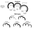 Foraminifera wall types.png 2032 × 1644; 388 кб
-
 Fresh-water rhizopods of North America (1879) (20174188464).jpg 2130 × 2852; 1,57 Мб
-
 Gastrocirrhus intermedius Lepsi, 1928 in Kahl 1932.png 533 × 783; 328 кб
-
 Gastrocirrhus stentoreus Bullington, 1940.png 516 × 742; 306 кб
-
 Genres Philaster et Philasterides in Kahl 1931.jpg 1566 × 1626; 393 кб
-
 Georg August Goldfuss Protozoa Infusoria Monades.jpg 462 × 360; 36 кб
-
 Gymnozoum viviparum in Meunier 1907 planche XX.png 533 × 678; 203 кб
-
 Hairs of a stramenopile.jpg 668 × 984; 229 кб
-
 Haptophrya gigantea Maupas, 1879 in Cépède 1910.jpg 1012 × 1334; 242 кб
-
 Haramonas cells.jpg 875 × 1000; 124 кб
-
 Helichona Plate, 1889 in Kahl 1935.jpg 1290 × 1035; 173 кб
-
 Helicoprorodon (Chanea) gigas (Kahl, 1933) Fauré-Fremiet, 1950.png 477 × 1232; 293 кб
-
 Heliophrya rotunda (Hentschel, 1916) Matthes, 1954.jpg 1041 × 2397; 897 кб
-
 Das Tierreich. 000, 1896 - Heliozoa (IA heliozoa00scha).pdf 941 × 1456, 38 старонак; 1,7 Мб
-
 Hemicycliostyla in Kahl 1932.png 756 × 845; 508 кб
-
 Hemimastix amphikineta.png 591 × 827; 97 кб
-
 Hemimastix amphikineta.svg 744 × 1052; 97 кб
-
 Hemiophrys fusidens & H. fossigera in Kahl 1931 et 1935.png 408 × 742; 202 кб
-
 Holosticha in Kahl 1932.png 1887 × 1596; 2,11 Мб
-
 Homalozoon caudatum in Kahl 1935.png 511 × 668; 210 кб
-
 Hypocoma parasitica in Bütschli, 1889.png 666 × 1286; 426 кб
-
 Hypotrichidium conicum Ilowaisky, 1921 in Kahl 1932.png 327 × 635; 152 кб
-
 Image from page 259 of "Fresh-water biology" (1918).jpg 276 × 362; 29 кб
-
 Intoshellina Maupasi Cépède 1910.jpg 1176 × 1941; 520 кб
-
 Isotricha prostoma et I. intestinalis in Kudo 1946.jpg 789 × 876; 231 кб
-
 John Hogg -- Primigenum or Protoctista.jpg 3305 × 1858; 534 кб
-
 Kerona in Muller 1786 Tab 33.jpg 2448 × 2414; 1,08 Мб
-
 Kerona in Muller 1786 Tab 34.jpg 2247 × 1419; 666 кб
-
-
 Keronopsis spectabilis Kahl, 1932 in Kahl 1932.png 438 × 877; 296 кб
-
 Kyste et enkystement d’Orchilophrya stellarum in Cépède 1910.jpg 926 × 1706; 313 кб
-
 Lacrymaria (31 schémas, 15 espèces) in Kahl 1930.png 1042 × 918; 826 кб
-
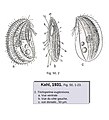
 Lagenophrys Stein, 1851 in Kahl 1935.png 1324 × 1468; 803 кб
-
 Lagynophrya sp & Kamburophrys gibba (Kahl, 1935) Foissner & Örtel, 2009.png 848 × 1013; 366 кб
-
 Lagynophrya sp. in Kahl 1930, 1935.png 984 × 1184; 841 кб
-
 Lagynus binucleatus Limin Jiang et al. 2021.png 1424 × 1378; 951 кб
-
 Lagynus in Maupas 1883.png 942 × 1080; 432 кб
-
 Lecanophrya drosera Kahl, 1934 (in A. Kahl 1934).png 595 × 591; 94 кб
-
 Leeuwenhoek-Little Animals Observed in Rain-Well-Sea.pdf 887 × 1429, 11 старонак; 1,73 Мб
-
 Leiotrocha Fabre-Domergue, 1888.jpg 2573 × 4318; 1,81 Мб
-
 Lembadion Perty, 1849 in Sheviakov, 1896.jpg 561 × 1297; 166 кб
-
 Lemboides Kahl, 1931 ou Paralembus Kahl, 1933.jpg 782 × 970; 136 кб
-
 Lembus Cohn, 1865.jpg 2312 × 1900; 705 кб
-
 Leptopharynx (ex Trichopelma) euglenivora Kahl, 1926.jpg 1280 × 1404; 254 кб
-
 Levels in complexity among different types of protist.jpg 742 × 469; 146 кб
-
 LifeCycleThraustochytrid.jpg 2343 × 2731; 1,12 Мб
-
 Lophophorina capronata Penard in Kahl 1931.png 534 × 641; 225 кб
-
 Loxophyllum armatum, setigerum et Meleagris in Bütschli 1887-1889.png 932 × 1317; 1,1 Мб
-
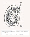
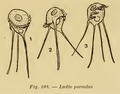 Ludio parvulus Penard, 1922.png 792 × 623; 159 кб
-
 Lycogala sp.jpg 2997 × 2421; 5,63 Мб
-
 Lynchella in Kahl 1935.png 658 × 599; 300 кб
-
 Malacophrys Kahl, 1926.jpg 668 × 1276; 208 кб
-
 Marine flagellates.jpg 570 × 125; 18 кб
-
 Maryna in A. Gruber 1879.png 702 × 1004; 286 кб
-
 Maupasella nova Cépède, 1910.jpg 2196 × 2964; 782 кб
-
 Metaradiophrya lumbrici Dujardin, 1841 in Obert & Vdacny 2019.jpg 818 × 1520; 408 кб
-
 Meteora sporadica.png 281 × 171; 6 кб
-
 Metromonas.jpg 91 × 100; 8 кб
-
 Microthorax et Hemicyclium in Kahl 1931.jpg 1263 × 2000; 582 кб
-
 Miliolid wall SEM.png 886 × 570; 470 кб
-
 Monocermonoides melolanthae.jpg 300 × 922; 37 кб
-
 Monosiga gracilis Kent = Codosiga gracilis (Kent, 1880) De Saedeleer, 1927.png 1125 × 1888; 1,18 Мб
-
 Multifasciculatum elegans Goodrich & Jahn, 1943.jpg 448 × 1102; 74 кб
-
 Nassula elegans Ehrenberg, 1833 in Sheviakov 1896 & Kahl 1931.jpg 890 × 1206; 303 кб
-

-
 Naviculoid diatom.jpg 1500 × 814; 136 кб
-
 Nicollella ctenodactyli et Collinella gundii in Buisson 1923.jpg 908 × 1232; 244 кб
-
 Normalized curves for flagellate and ciliate swimming speeds.jpg 1500 × 532; 74 кб
-
 Nutomonas@SciELO.jpg 1212 × 898; 69 кб
-
 Nyctotherus Leidy, 1849 in R.P. Hall 1953.png 1516 × 1512; 855 кб
-
 Occultammina sp.png 788 × 514; 434 кб
-
 Odontochlamys in Certes 1891.png 1088 × 1752; 390 кб
-
 Onychodromus grandis in Maupas 1888.png 1154 × 1486; 1,65 Мб
-
 Onychodromus grandis in Maupas 1889.jpg 3720 × 5928; 3,18 Мб
-
 Onychodromus grandis Stein, 1859 in Kahl 1932.png 311 × 820; 220 кб
-
 Opercularia Goldfuss, 1820 in Saville-Kent, 1888.jpg 1980 × 3091; 1,59 Мб
-
 Ophiocytium.jpg 1304 × 1000; 142 кб
-
 Ophrydium versatile (Müller, 1786) Bory de St.Vincent, 1826 in Saville-Kent 1882.jpg 2008 × 2568; 1,37 Мб
-

-
 Ophryoglena Ehrenberg, 1831.jpg 1779 × 2460; 999 кб


_Jankowski,_1978.png)


_(1910)_(17950796051)-9%2B10%2B11.jpg)


_in_Journal_de_Microgr._1889.png)


_opheliae_Awerinzew,_1908.jpg)


_in_Kahl_1931_et_1935.png)


_Perty,_1849.jpg)


_punctatus_Kahl,_1933.jpg)
_C%C3%A9p%C3%A8de,_1910.jpg)


_Raabe,_1936.png)

.jpg)


_(20723576510).jpg)
_(20918951511).jpg)
_043_Wassertropfen_aus_gestandenem_Wasser.png)
_des_golfes_von_Neapel,_und_der_angrenzenden_meeresabschnitte_(1885)_(20308769293).jpg)
_des_golfes_von_Neapel,_und_der_angrenzenden_meeresabschnitte_(1885)_(20741742510).jpg)
_des_golfes_von_Neapel,_und_der_angrenzenden_meeresabschnitte_(1885)_(20743061889).jpg)
_des_golfes_von_Neapel,_und_der_angrenzenden_meeresabschnitte_(1885)_(20920085202).jpg)
_des_golfes_von_Neapel,_und_der_angrenzenden_meeresabschnitte_(1885)_(20937161351).jpg)
_-_eine_Monographie_(1887)_(20754175700).jpg)
_Jankowski,_1967.jpg)


_(20586649844).jpg)
_(21021175860).jpg)
_(21021178970).jpg)
_(21022467779).jpg)
_(21183081446).jpg)
_(21183086586).jpg)
_(21217233731).jpg)
_(21217236511).jpg)


_Kahl,_1930.jpg)


_Crawley,_1923_in_Kudo_1946.jpg)


_(20174188464).jpg)


_Matthes,_1954.jpg)


.jpg)


_in_B%C3%BCtschli_1889.png)


_in_Kahl_1930.png)

_Foissner_%26_%C3%96rtel,_2009.png)


.png)


_euglenivora_Kahl,_1926.jpg)


_De_Saedeleer,_1927.png)

_(5974464293).jpg)


_Bory_de_St.Vincent,_1826_in_Saville-Kent_1882.jpg)


_gigas_(Kahl,_1933)_Faur%C3%A9-Fremiet,_1950.png)