File:C elegans male.svg

Original file (SVG file, nominally 1,408 × 2,250 pixels, file size: 635 KB)
Captions
Captions
Summary[edit]
| Description |
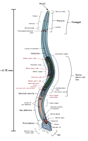
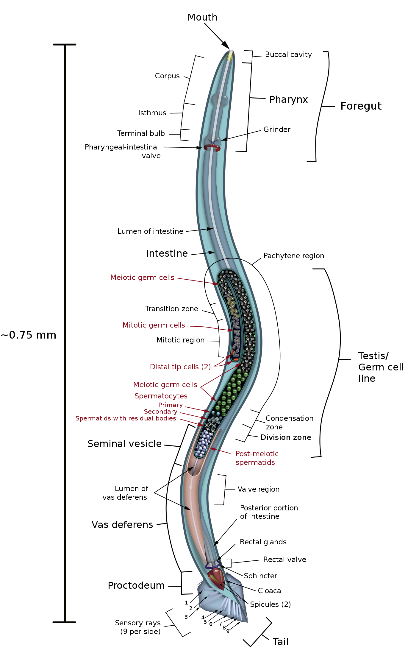
English: Anatomical drawing of a male C. elegans nematode with emphasis on the reproductive system. Note that the actual worm is colorless and transparent. Also note that the raw SVG version of the image will not render correctly in Firefox 23.0.1 or earlier (possibly later as well) due to known Mozilla program bug #376027 but should do so in any other browser. |
| Date | |
| Source | Own work |
| Author | KDS444 |
| Other versions |
[]
|
| SVG development | This diagram was created with Adobe Illustrator. This diagram uses embedded text that can be easily translated using a text editor. |


|
This image was selected as picture of the day on Wikimedia Commons for 21 October 2013. It was captioned as follows: English: Anatomical drawing of a male C. elegans nematode with emphasis on the reproductive system. Other languages:
English: Anatomical drawing of a male C. elegans nematode with emphasis on the reproductive system. Italiano: Disegno anatomico di un esemplare maschio di verme nematode Caenorhabditis elegans con enfasi sul sistema di riproduzione. Magyar: A fonálférgek (Nematoda) törzsébe és az érzékpálcások (Secernentea) osztályának Caenorhabditis nemébe tartozó Caenorhabditis elegans reproduktív szervrendszerét kiemelő anatómiai rajz Nederlands: Anatomische tekening van een mannelijke Caenorhabditis elegans met nadruk op het voortplantsingssysteem. Русский: Анатомия самца круглого червя Caenorhabditis elegans. 日本語: 線虫の一種、C. elegans (カエノラブディティス・エレガンス, 和名なし, 通称:シー・エレガンス)♂の解剖図。 生殖系の臓器群を強調した図。 中文: 秀丽隐杆线虫的解刨图,着重强调了生殖系统。 |
Sources drawn upon in the creation of this image include the following, among others:
- ↑ Maine, Elanor M. (2010), “Meiotic silencing in Caenorhabditis elegans”, in International Review of Cell and Molecular Biology[1], volume 282, Elsevier, pages 91-134
- ↑ Hansen, David (2012-04-10). The Hansen Lab - Research. University of Calgary. Archived from the original on 2015-07-17.
- ↑ Zarkower, David (2006), “Somatic sex determination”, in Wormbook[2], The C. elegans Research Community, DOI:
- ↑ Lints, R.; Hall, D.H. (2009), “Male reproductive system, general description”, in WormAtlas[3], DOI:
- ↑ Lints, R.; Hall, D.H. (2009), “Male reproductive system, somatic gonad”, in WormAtlas[4], DOI:
- ↑ Lints, R.; Hall, D.H. (2009), “Male reproductive system, germ line”, in WormAtlas[5], DOI:
- ↑ Lints, R.; Hall, D.H. (2009), “Male reproductive system, proctodeum”, in WormAtlas[6], DOI:
Licensing[edit]
- You are free:
- to share – to copy, distribute and transmit the work
- to remix – to adapt the work
- Under the following conditions:
- attribution – You must give appropriate credit, provide a link to the license, and indicate if changes were made. You may do so in any reasonable manner, but not in any way that suggests the licensor endorses you or your use.
- share alike – If you remix, transform, or build upon the material, you must distribute your contributions under the same or compatible license as the original.
Illustration license notice[edit]
|
|
This image is NOT in the PUBLIC DOMAIN. If you wish to use this image on a different web site or publication you are obliged to provide following details along with the image:
|
File history
Click on a date/time to view the file as it appeared at that time.
| Date/Time | Thumbnail | Dimensions | User | Comment | |
|---|---|---|---|---|---|
| current | 22:08, 28 August 2017 | | 1,408 × 2,250 (635 KB) | KDS4444 (talk | contribs) | raster gone |
| 20:29, 28 August 2017 |  | 1,684 × 2,268 (579 KB) | KDS4444 (talk | contribs) | fixing w3c errors | |
| 04:56, 8 May 2015 |  | 1,684 × 2,559 (707 KB) | KDS4444 (talk | contribs) | minor improvements | |
| 13:46, 28 May 2014 |  | 3,072 × 4,976 (741 KB) | KDS444 (talk | contribs) | even better | |
| 20:21, 5 September 2013 |  | 1,684 × 2,268 (571 KB) | KDS444 (talk | contribs) | for crying out loud | |
| 20:18, 5 September 2013 |  | 1,684 × 2,268 (571 KB) | KDS444 (talk | contribs) | I effing hate Myriad Pro. | |
| 20:15, 5 September 2013 |  | 1,684 × 2,268 (571 KB) | KDS444 (talk | contribs) | Have corrected white "balloon" around mouth, and am taking a shot at fixing the font alignment/ size problem in the middle left portion of the image. | |
| 19:35, 5 September 2013 |  | 1,684 × 2,268 (571 KB) | KDS444 (talk | contribs) | Improved appearance of meiotics | |
| 01:13, 2 September 2013 |  | 1,444 × 2,210 (666 KB) | KDS444 (talk | contribs) | Softening of contrast in early meiotic germ cells | |
| 18:25, 29 August 2013 |  | 1,444 × 2,210 (666 KB) | KDS444 (talk | contribs) | sensory ray openings no longer boxes |
You cannot overwrite this file.
File usage on Commons
The following 53 pages use this file:
- SVG animal diagrams
- User:KDS4444
- User:KDS4444/Profile
- User:Miya/sandbox/FP/2013/Galleries/Table
- User talk:KDS444
- Commons:Bu proje sayfasının diğer dil sürümleri
- Commons:Conhece os nossos ilustradores
- Commons:Conoce a nuestros ilustradores
- Commons:Descobrètz nòstres illustrators
- Commons:Découvrez nos illustrateurs
- Commons:Featured picture candidates/File:C elegans male.svg
- Commons:Featured picture candidates/Log/September 2013
- Commons:Featured pictures/Non-photographic media/Computer-generated
- Commons:Featured pictures/chronological/2013-B
- Commons:Meet our illustrators
- Commons:Meet our illustrators/People
- Commons:Picture of the Year/2013/Candidates
- Commons:Picture of the Year/2013/Galleries/Table
- Commons:Picture of the Year/2013/R1/Gallery/2013-B
- Commons:Picture of the Year/2013/R1/Gallery/ALL
- Commons:Picture of the Year/2013/R1/Gallery/M09
- Commons:Picture of the Year/2013/R1/Gallery/Maps
- Commons:Picture of the Year/2013/R1/Results/Candidates
- Commons:Picture of the Year/2013/R1/v/C elegans male.svg
- Commons:Poznaj naszych grafików
- Commons:Scopri i nostri illustratori
- Commons:Scummigghia ê nostri llustratura
- Commons:Unsere Illustratoren stellen sich vor
- Commons:Unseri Illustratore stelle sich vor
- Commons:Upoznajte naše ilustratore
- Commons:Çizerlerimizle tanışın
- Commons:Запознајте ги нашите илустратори
- Commons:Знайомтесь з нашими ілюстраторами
- Commons:Знакомьтесь с нашими иллюстраторами
- Commons:قابل رسامينا
- Commons:私達のイラストレータをご紹介します
- Commons:结识我们的插图师
- Commons:우리들의 삽화가를 만나보세요
- File:Anatomie d'un nématode masculin-fr.svg
- File:Anatomía de un nematodo macho-es.svg
- File:C elegans male-gl.svg
- File:C elegans male.svg
- Template:Other versions/C elegans male
- Template:Potd/2013-10
- Template:Potd/2013-10-21
- Template:Potd/2013-10-21 (en)
- Template:Potd/2013-10-21 (hu)
- Template:Potd/2013-10-21 (it)
- Template:Potd/2013-10-21 (ja)
- Template:Potd/2013-10-21 (nl)
- Template:Potd/2013-10-21 (ru)
- Template:Potd/2013-10-21 (zh-hans)
- Template:Potd/2013-10 (zh-hans)
File usage on other wikis
The following other wikis use this file:
- Usage on arz.wikipedia.org
- Usage on as.wikipedia.org
- Usage on be-tarask.wikipedia.org
- Usage on bn.wikipedia.org
- Usage on bs.wikipedia.org
- Usage on ca.wikipedia.org
- Usage on ca.wikiquote.org
- Usage on crh.wikipedia.org
- Usage on cv.wikipedia.org
- Usage on cy.wikipedia.org
- Usage on en.wikipedia.org
- Caenorhabditis elegans
- Talk:Caenorhabditis elegans
- Wikipedia:Featured pictures/Animals/Others
- Nematode
- Wikipedia:Featured pictures thumbs/36
- Wikipedia:Featured picture candidates/Male C. elegans anatomy
- Wikipedia:Featured picture candidates/September-2013
- Wikipedia:Picture of the day/October 2013
- Template:POTD/2013-10-13
- Wikipedia:Main Page history/2013 October 13
- User talk:KDS4444/Archive 1
- Usage on eu.wikipedia.org
- Usage on hak.wikipedia.org
- Usage on ha.wikipedia.org
- Usage on hu.wikipedia.org
- Usage on ka.wikipedia.org
- Usage on ko.wikipedia.org
- Usage on krc.wikipedia.org
- Usage on lbe.wikipedia.org
- Usage on lfn.wikipedia.org
- Usage on ms.wiktionary.org
- Usage on nl.wikipedia.org
- Usage on nn.wikipedia.org
- Usage on os.wikipedia.org
- Usage on pt.wikipedia.org
- Usage on ru.wikipedia.org
- Usage on ru.wikinews.org
- Usage on sah.wikipedia.org
- Usage on sh.wikipedia.org
- Usage on species.wikimedia.org
- Usage on sr.wikipedia.org
- Usage on ta.wikipedia.org
View more global usage of this file.