File:FIPCytology2.jpg
FIPCytology2.jpg (350 × 234 pixels, file size: 40 KB, MIME type: image/jpeg)
Captions
Captions
Summary[edit]
| Description |
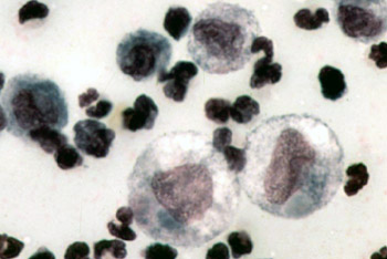
English: Color micrograph of the cytology of FIP-induced effusion. Magnification not specified; estimated to be 1000x.
Original caption: "The cytology of FIP effusion usually contains neutrophils, macrophages and lymphocytes." Image from "Feline Infectious Peritonitis: An Overview of Disease Transmission, Pathogenesis, Signs and Treatment With Emphasis on Diagnosis" ([1]) Clinical Pathology Clerkship Program |
| Date | 30 September 2005 (original upload date) |
| Source | Transferred from en.wikipedia to Commons. |
| Author | The original uploader was Bk0 at English Wikipedia. |
Licensing[edit]

|
The copyright holder of this file allows anyone to use it for any purpose, provided that the copyright holder is properly attributed. Redistribution, derivative work, commercial use, and all other use is permitted. |
|
|
Original upload log[edit]
- 2005-09-30 00:14 Bk0 350×234×8 (40687 bytes) Color micrograph of the cytology of [[Feline infectious peritonitis|FIP]]-induced effusion. Magnification not specified; estimated to be 1000x. Original caption: "The cytology of FIP effusion usually contains neutrophils, macrophages and lymphocytes." I
File history
Click on a date/time to view the file as it appeared at that time.
| Date/Time | Thumbnail | Dimensions | User | Comment | |
|---|---|---|---|---|---|
| current | 18:53, 29 December 2007 | | 350 × 234 (40 KB) | Euthygenes (talk | contribs) | {{Information |Description={{en|Color micrograph of the cytology of FIP-induced effusion. Magnification not specified; estimated to be 1000x. Original caption: "The cytology of FIP effusion usually contains neutrophi |
You cannot overwrite this file.
File usage on Commons
There are no pages that use this file.
File usage on other wikis
The following other wikis use this file:
- Usage on el.wikipedia.org
- Usage on en.wikipedia.org
- Usage on et.wikipedia.org
- Usage on fr.wikipedia.org
- Usage on hu.wikipedia.org
- Usage on ko.wikipedia.org
- Usage on tr.wikipedia.org
- Usage on zh.wikipedia.org