File:Human skeletal muscle tissue 2 - TEM.jpg

Original file (2,191 × 1,630 pixels, file size: 1.24 MB, MIME type: image/jpeg)
Captions
Captions
Summary[edit]
| Description |
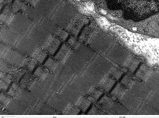
Transmission electron microscope image of a thin longitudinal section cut through an area of human skeletal muscle tissue. Image shows several myofibrils, each with distinct banding pattern of individual sarcomeres. Image of muscle sarcomeres shows distinct banding pattern: the darker bands are called A bands(the A band includes a lighter central zone, called the H band), and the lighter bands are called I bands. Each I band is bisected by a dark transverse line called the Z-line). Paired mitochondria are on either side of the electron opaque Z-line. The Z-Line marks the longitudinal extent of a sarcomere unit. JEOL 100CX TEM |
| Source | |
| Author | Louisa Howard |
| Permission (Reusing this file) |
PD |
Licensing[edit]
| This work has been released into the public domain by its author, Louisa Howard. This applies worldwide. In some countries this may not be legally possible; if so: Louisa Howard grants anyone the right to use this work for any purpose, without any conditions, unless such conditions are required by law.
|
File history
Click on a date/time to view the file as it appeared at that time.
| Date/Time | Thumbnail | Dimensions | User | Comment | |
|---|---|---|---|---|---|
| current | 14:58, 7 October 2006 | | 2,191 × 1,630 (1.24 MB) | Patho (talk | contribs) | {{Information |Description= Transmission electron microscope image of a thin longitudinal section cut through an area of human skeletal muscle tissue. Image shows several myofibrils, each with distinct banding pattern of individual sarcomeres. Image of |
You cannot overwrite this file.
File usage on Commons
There are no pages that use this file.
File usage on other wikis
The following other wikis use this file:
- Usage on de.wikibooks.org
- Usage on fr.wikipedia.org
- Usage on hu.wikipedia.org
- Usage on ko.wikipedia.org
- Usage on lt.wikipedia.org