File:Lung tissue during legionellosis.jpg
Jump to navigation
Jump to search

Size of this preview: 800 × 531 pixels. Other resolutions: 320 × 213 pixels | 640 × 425 pixels | 1,024 × 680 pixels | 1,280 × 850 pixels | 1,814 × 1,205 pixels.
Original file (1,814 × 1,205 pixels, file size: 1.99 MB, MIME type: image/jpeg)
Captions
Captions
Add a one-line explanation of what this file represents
Summary[edit]
| Description |
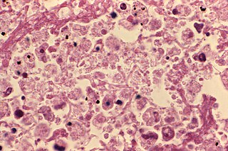
Русский: На данной микроскопической картине представлена ткань легкого человека, больного легионеллезной пневмонией. Окраска гематоксилин-эозином. Legionella pneumophila грам-отрицательные, видны в виде красных палочек. Ткань легкого инфильтрирована большим количеством лейкоцитов.
English: This micrograph depicted details seen in a lung tissue specimen from a Knoxville patient with fatal pneumonia due to Legionnaires’ disease. The tissue was stained using hematoxylin-eosin (H&E) stain. Legionella pneumophila are Gram-negative bacteria. Using H&E stain, these organisms, if present in the specimen, would stain a pink or red color. Note how the alveolar spaces are so congested with a leukocytic infiltrate in response to the infection. |
|||
| Date | ||||
| Source |
|
|||
| Author | CDC/ William Cherry | |||
| Permission (Reusing this file) |
|
File history
Click on a date/time to view the file as it appeared at that time.
| Date/Time | Thumbnail | Dimensions | User | Comment | |
|---|---|---|---|---|---|
| current | 20:12, 30 November 2010 | | 1,814 × 1,205 (1.99 MB) | Masur (talk | contribs) | orig. res. same source |
| 20:22, 30 May 2010 |  | 700 × 464 (62 KB) | Yartsew (talk | contribs) | {{Information |Description={{ru|1=На данной микроскопической картине представлена ткань легкого человека, больного легионеллезной пневмонией. Окраска г� |
You cannot overwrite this file.
File usage on Commons
There are no pages that use this file.
File usage on other wikis
The following other wikis use this file:
- Usage on es.wikipedia.org
- Usage on ru.wikipedia.org
- Usage on sr.wikipedia.org
- Usage on uk.wikipedia.org