File:RNA-in-situ-dark-field.jpg
From Wikimedia Commons, the free media repository
Jump to navigation
Jump to search
RNA-in-situ-dark-field.jpg (364 × 198 pixels, file size: 62 KB, MIME type: image/jpeg)
File information
Structured data
Captions
Captions
Add a one-line explanation of what this file represents
| DescriptionRNA-in-situ-dark-field.jpg |
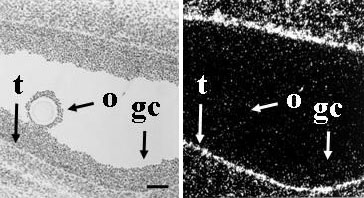
English: Tissue section with radioactive RNA in situ hybridization followed by coating with autoradiographic emulsion, development of silver grains and hematoxylin staining. See Methods section of original paper for details.
From the original figure legend: Localization of expression of mRNA encoding TGF-βs in ovine ovaries. Panels a and b contain corresponding light field and dark field views of a type 5 follicle from an adult ewe hybridized to TGF-β1 antisense RNA. Silver grains indicating hybridization of the TGF-β1 antisense RNA are observed concentrated in thecal (t) cells close to the basement membrane with no specific hybridization observed in either the granulosa cells (gc) or oocyte (o). Scale bar equals approximately 100 μm.Deutsch: Gewebeschnitt nach radioaktiver RNA in situ Hybridisierung und Nachweis der Radioaktivität durch Entstehung von Silberkörnern. Bei Hellfeld-Mikroskopie (links) sind diese kaum zu sehen, bei Dunkelfeldmikroskopie (rechts) treten sie dagegen deutlich hervor. |
| Date | |
| Source | Reproductive Biology and Endocrinology 2004, 2:78 doi:10.1186/1477-7827-2-78. Figure 1. Online |
| Author | Jennifer L Juengel, Adrian H Bibby, Karen L Reader, Stan Lun, Laurel D Quirke, Lisa J Haydon and Kenneth P McNatty |
| Permission (Reusing this file) |
This file is licensed under the Creative Commons Attribution 2.0 Generic license.
|
File history
Click on a date/time to view the file as it appeared at that time.
| Date/Time | Thumbnail | Dimensions | User | Comment | |
|---|---|---|---|---|---|
| current | 14:20, 20 May 2012 |  | 364 × 198 (62 KB) | Dietzel65 (talk | contribs) | {{Information |Description ={{en|1=Tissue section with radioactive RNA in situ hybridization followed by coating with autoradiographic emulsion, development of silver grains and hematoxylin staining. See Methods section of original paper for details... |
You cannot overwrite this file.
File usage on Commons
The following page uses this file:
File usage on other wikis
The following other wikis use this file:
- Usage on de.wikipedia.org
- Usage on fr.wikipedia.org
- Usage on nl.wikipedia.org
Metadata
This file contains additional information such as Exif metadata which may have been added by the digital camera, scanner, or software program used to create or digitize it. If the file has been modified from its original state, some details such as the timestamp may not fully reflect those of the original file. The timestamp is only as accurate as the clock in the camera, and it may be completely wrong.
| Orientation | Normal |
|---|---|
| Horizontal resolution | 72 dpi |
| Vertical resolution | 72 dpi |
| Software used | Adobe Photoshop CS2 Windows |
| File change date and time | 16:05, 20 May 2012 |
| Color space | sRGB |
| Image width | 364 px |
| Image height | 198 px |
| Date and time of digitizing | 18:05, 20 May 2012 |
| Date metadata was last modified | 18:05, 20 May 2012 |
Structured data
Items portrayed in this file
depicts
Hidden category:
