File:Thyroid papillary carcinoma histopathology (4).jpg
Jump to navigation
Jump to search
No higher resolution available.
Thyroid_papillary_carcinoma_histopathology_(4).jpg (500 × 376 pixels, file size: 59 KB, MIME type: image/jpeg)
Captions
Captions
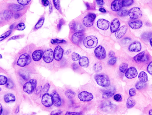
Histopatholgical image of papillary carcinoma of the thyroid gland
Summary[edit]
.jpg&action=edit§ion=1)
| Description |
English: Histopatholgical image of papillary carcinoma of the thyroid gland obtained by a total thyroidectomy. Hematoxylin and eosin stain. Another version of an accompanying file "Thyroid_papillary_carcinoma_histopathology_(3).jpg". |
| Source | No machine-readable source provided. Own work assumed (based on copyright claims). |
| Author | No machine-readable author provided. KGH assumed (based on copyright claims). |
Licensing[edit]
.jpg&action=edit§ion=2)
I, the copyright holder of this work, hereby publish it under the following licenses:

|
Permission is granted to copy, distribute and/or modify this document under the terms of the GNU Free Documentation License, Version 1.2 or any later version published by the Free Software Foundation; with no Invariant Sections, no Front-Cover Texts, and no Back-Cover Texts. A copy of the license is included in the section entitled GNU Free Documentation License. |
| This file is licensed under the Creative Commons Attribution-Share Alike 3.0 Unported license. | ||
| ||
| This licensing tag was added to this file as part of the GFDL licensing update. |
You may select the license of your choice.
| Annotations | This image is annotated: View the annotations at Commons |
.jpg)
File history
Click on a date/time to view the file as it appeared at that time.
| Date/Time | Thumbnail | Dimensions | User | Comment | |
|---|---|---|---|---|---|
| current | 14:55, 8 January 2006 | | 500 × 376 (59 KB) | KGH (talk | contribs) | Histopatholgical image of papillary carcinoma of the thyroid gland obtained by a total thyroidectomy. Hematoxylin and eosin stain. Another version of an accompanying file "Thyroid_papillary_carcinoma_histopathology_(3).jpg". |
You cannot overwrite this file.
File usage on Commons
The following page uses this file:
File usage on other wikis
The following other wikis use this file:
- Usage on ar.wikipedia.org
- Usage on bn.wikipedia.org
- Usage on bs.wikipedia.org
- Usage on ca.wikipedia.org
- Usage on en.wikipedia.org
- Usage on es.wikipedia.org
- Usage on fa.wikipedia.org
- Usage on gl.wikipedia.org
- Usage on hy.wikipedia.org
- Usage on it.wikipedia.org
- Usage on ja.wikipedia.org
- Usage on ko.wikipedia.org
- Usage on new.wikipedia.org
- Usage on or.wikipedia.org
- Usage on pt.wikipedia.org
- Usage on ru.wikipedia.org
- Usage on sh.wikipedia.org
- Usage on sk.wikipedia.org
- Usage on sr.wikipedia.org
- Usage on sw.wikipedia.org
- Usage on tr.wikipedia.org
- Usage on zh.wikipedia.org
.jpg&oldid=743390919)