Horse hoof
Here some pictures of Horse hoof.
Pictures[编辑]
-
 Fig. 1
 -
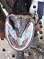 Fig 2
Fig 2 -
 Fig 3
Fig 3 -
 Fig 4
Fig 4 -
 Fig 5
Fig 5 -
 Fig 6
Fig 6


Here the Italian version of a legend for Pict 2:
1- Perioplio del tallone, 2-Glomo, 3-Fettone, 4-Lacuna centrale, 5-Lacuna collaterale, 6-Tallone, 7-Barra, 8-Angolo di inflessione, 9-Muraglia pigmentata (strato esterno), 10-Muraglia non pigmentata (strato interno), 11-Linea bianca, 12-Punta del fettone, 13-Suola, 14-Punta dello zoccolo, 15-Guida per la misura della larghezza (linea azzurra tratteggiata), 16-Quarto, 17-Guida per la misura della lunghezza (linea azzurra tratteggiata)
Here the Italian version of a legend for Pict 3: 1-Corona 2-Muraglia 3-Punta 4-Quarto 5-Tallone 6-Glomo 7-Piccolo pastorale (2° falange)
Here an English legend (please correct any mistake!) for Pict 2: 1- Heel perioplium, 2-Bulb, 3-Frog, 4-Frog cleft, 5-Lateral groove, 6-Heel, 7-Bar, 8-Seat-of-corn, 9-Pigmented walls 10-Water line, 11-White line, 12-Apex of the frog, 13-Sole, 14-Toe, 15-How to measure hoof width (blu dotted line), 16-Quarter, 17-How to measrure length (blue dotted line)
And for Pict 3: 1-Coronary band 2-Walls 3-Toe 4-Quarter 5-Heel 6-Bulb 7-Small pastern (P2 = Phalanx 2)
Pict. 4: Severe laminitis (founder); hoof specimen, sagittal section Pict. 5: Bare hooves prints Pict. 6: Shoeing in progress
Drawings[编辑]


The second and the third drawing show a shod hoof; the former when shoe has just nailed on, the latter some weeks after. Note that when the walls grow up, there is no more contact between le sole and the shoe; the gravity stress from the bone column is only sustained by walls laminar connections, without any solar/frog support. In the opinion of Pete Ramey [1], a resulting, slow downward movement of bony column into the hoof capsule results, as showed by "long walls" and apparent "upward coronet displacing" (sometimes, with conflict between the walls and the P2-P3 joint) in middle-aged or older shod horses.
See images for a brief legend.