Category:Cells
Vai alla navigazione
Vai alla ricerca
unità fondamentale degli organismi viventi  | |||||
| Carica un file multimediale | |||||
| Istanza di |
| ||||
|---|---|---|---|---|---|
| Sottoclasse di |
| ||||
| Parte di |
| ||||
| Consiste di |
| ||||
| Distinto da | |||||
| |||||
Sottocategorie
Questa categoria contiene le 4 sottocategorie indicate di seguito, su un totale di 4.
File nella categoria "Cells"
Questa categoria contiene 200 file, indicati di seguito, su un totale di 373.
(pagina precedente) (pagina successiva)-
 12985 2005 Article 81 Fig2 HTML.jpg 1 200 × 824; 104 KB
-
 201012 CHOcells.png 663 × 570; 263 KB
-
 201012 S2 cells.png 500 × 414; 291 KB
-
 201012 Vero.png 408 × 433; 188 KB
-
 201101 HEK293.png 781 × 605; 323 KB
-
 201101 NIH3T3.png 800 × 600; 122 KB
-
 201101 Sf9.png 800 × 600; 545 KB
-
 201102 adipocyte.png 524 × 464; 182 KB
-
 201102 astrocyte.png 458 × 440; 75 KB
-
 201102 chondrocyte.png 640 × 445; 182 KB
-
 201102 mammary epithelial cell.png 318 × 346; 62 KB
-
 201102 neuron.png 582 × 634; 68 KB
-
 201102 stem cell.png 260 × 291; 70 KB
-
 201108 budding yeast.png 329 × 209; 22 KB
-
 201304 blastcyst.png 500 × 375; 113 KB
-
 201304 fertilized egg.png 500 × 375; 86 KB
-
 201304 gastrula.png 500 × 375; 101 KB
-
 201304 sperm.png 500 × 375; 14 KB
-
 201308 platelet.png 500 × 375; 34 KB
-
 201904 C2C12.svg 512 × 410; 1,32 MB
-
 202011 Cardiac Myocytes.svg 512 × 512; 1,05 MB
-
 202312 granulocytes.svg 400 × 400; 3 KB
-
 202312 oenocytoide.svg 400 × 400; 2 KB
-
 202312 plasma cell.svg 400 × 400; 1 KB
-
 202312 prohemocyte.svg 400 × 400; 949 byte
-
 202312 spherule cell.svg 400 × 400; 4 KB
-
 208031 EPFL David Suter Sox2.jpg 1 304 × 734; 52 KB
-
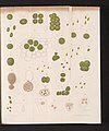 32 voorstellingen van cellen, RP-F-2001-7-960-9.jpg 2 944 × 3 522; 960 KB
-
 405 Modes of Secretion by Glands Merocine.png 1 254 × 579; 268 KB
-
 43 voorstellingen van cellen, RP-F-2001-7-960-7.jpg 4 098 × 5 052; 2,37 MB
-
 55 voorstellingen van cellen, RP-F-2001-7-960-8.jpg 4 184 × 5 076; 2,17 MB
-
 A Cell Post Plasmolysis.jpg 960 × 720; 94 KB
-
 Animal cells SwissBioPics DL20221120.svg 512 × 430; 371 KB
-
 Anisocytosis 2.jpg 1 280 × 1 024; 72 KB
-
 Apoptosisgif.gif 1 090 × 970; 232 KB
-
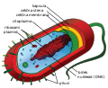 Average prokaryote cell- sl.svg 494 × 402; 145 KB
-
 Bacill.jpg 3 821 × 4 434; 558 KB
-
 Bernard-S.-Marasa-etal-2010-WI38.png 615 × 372; 194 KB
-

-
-
 Blood Anemia.jpg 1 280 × 720; 187 KB
-
 Camillo Golgi, Golgi cell Type I. Wellcome L0002017.jpg 1 184 × 1 522; 814 KB
-
 Cardiac Stem Cell Differentiation.png 512 × 444; 64 KB
-
 CardiacMuscle - longtitudinal.jpg 640 × 480; 188 KB
-
 Cell biology illustration by Franz Wagner, Wellcome L0033028.jpg 4 551 × 3 476; 5,5 MB
-
 Cell metabolism and color.jpg 3 888 × 2 592; 1,21 MB
-
 Cell mosaic.png 1 106 × 1 100; 1,17 MB
-
 Cell Scaffolding (12753438075).jpg 612 × 746; 180 KB
-
 Cell tower with sun set.jpg 1 080 × 1 080; 49 KB
-
 CellAdhesion.jpg 1 344 × 1 024; 203 KB
-
 Cells 001.png 734 × 1 077; 581 KB
-
 Cells 002.png 734 × 1 077; 524 KB
-
 Cells 003.png 1 091 × 1 079; 347 KB
-
 Cells 004.png 1 084 × 1 057; 346 KB
-
 Cells 005.png 1 074 × 1 194; 1,44 MB
-
 Cells 006.png 1 218 × 1 030; 1,13 MB
-
 Cells 007.png 1 162 × 1 055; 1,08 MB
-
 Cells 008.png 1 208 × 1 048; 1,15 MB
-
 Cells 009.png 1 184 × 1 055; 1,13 MB
-
 Cells 010.png 1 248 × 1 048; 1,15 MB
-
 Cells 011.png 836 × 806; 835 KB
-
 Cells 012.png 836 × 806; 901 KB
-
 Cells 013.png 966 × 866; 1,01 MB
-
 Cells 014.png 966 × 866; 1,05 MB
-
 Cells 015.png 738 × 573; 576 KB
-
 Cells 016.png 738 × 573; 584 KB
-
 Cells 017.png 791 × 475; 604 KB
-
 Cells 018.png 791 × 475; 609 KB
-
 Cells 019.png 692 × 730; 563 KB
-
 Cells 020.png 705 × 693; 357 KB
-
 Cells 021.png 889 × 981; 972 KB
-
 Cells 022.png 925 × 871; 1,05 MB
-
 Cells 023.png 740 × 731; 678 KB
-
 Cells 024.png 728 × 718; 610 KB
-
 Cells 025.png 752 × 754; 770 KB
-
 Cells 026.png 765 × 1 004; 662 KB
-
 Cells 027.png 765 × 1 004; 650 KB
-
 Cells 028.png 1 047 × 460; 311 KB
-
 Cells 029.png 1 043 × 459; 411 KB
-
 Cells 030.png 1 168 × 683; 543 KB
-
 Cells 031.png 1 168 × 683; 537 KB
-
 Cells 032.png 793 × 465; 472 KB
-
 Cells 033.png 793 × 465; 465 KB
-
 Cells 034.png 865 × 654; 363 KB
-
 Cells 035.png 865 × 654; 359 KB
-
 Cells 036.png 1 057 × 568; 471 KB
-
 Cells 037.png 1 057 × 568; 464 KB
-
 Cells 038.png 864 × 666; 513 KB
-
 Cells 039.png 864 × 666; 478 KB
-
 Cells 040.png 783 × 449; 423 KB
-
 Cells 041.png 783 × 449; 428 KB
-
 Cells 042.png 733 × 635; 483 KB
-
 Cells 043.png 736 × 724; 532 KB
-
 Cells 044.png 692 × 590; 461 KB
-
 Cells 045.png 737 × 708; 550 KB
-
 Cells 047.png 721 × 771; 579 KB
-
 Cells 048.png 707 × 586; 422 KB
-
 Cells 049.png 648 × 594; 398 KB
-
 Cells 050.png 759 × 697; 543 KB
-
 Cells 051.png 667 × 719; 524 KB
-
 Cells 052.png 722 × 717; 522 KB
-
 Cells 053.png 707 × 749; 596 KB
-
 Cells 054.png 724 × 749; 577 KB
-
 Cells 055.png 715 × 779; 560 KB
-
 Cells 056.png 605 × 659; 416 KB
-
 Cells 057.png 764 × 676; 454 KB
-
 Cells 058.png 622 × 668; 343 KB
-
 Cells 059.png 723 × 673; 422 KB
-
 Cells 060.png 691 × 647; 388 KB
-
 Cells 061.png 658 × 610; 348 KB
-
 Cells 062.png 765 × 725; 527 KB
-
 Cells 063.png 665 × 623; 434 KB
-
 Cells 064.png 695 × 600; 443 KB
-
 Cells 065.png 721 × 624; 425 KB
-
 Cells 066.png 770 × 709; 525 KB
-
 Cells 067.png 755 × 670; 483 KB
-
 Cells 068.png 656 × 708; 429 KB
-
 Cells 069.png 716 × 583; 401 KB
-
 Cells 070.png 636 × 592; 395 KB
-
 Cells 071.png 755 × 697; 481 KB
-
 Cells 072.png 764 × 676; 379 KB
-
 Cells 073.png 691 × 647; 342 KB
-
 Cells 074.png 658 × 610; 315 KB
-
 Cells 075.png 622 × 668; 320 KB
-
 Cells 076.png 723 × 673; 352 KB
-
 Cells 077.png 715 × 779; 680 KB
-
 Cells 078.png 722 × 717; 626 KB
-
 Cells 079.png 691 × 647; 542 KB
-
 Cells 080.png 622 × 668; 521 KB
-
 Cells 081.png 658 × 610; 517 KB
-
 Cells 082.png 764 × 676; 593 KB
-
 Cells 083.png 755 × 697; 586 KB
-
 Cells 084.png 636 × 592; 471 KB
-
 Cells 085.png 695 × 600; 516 KB
-
 Cells 086.png 724 × 749; 643 KB
-
 Cells 087.png 1 217 × 272; 407 KB
-
 Cells 088.png 1 217 × 272; 400 KB
-
 Cells 089.png 1 349 × 223; 315 KB
-
 Cells 090.png 1 349 × 223; 311 KB
-
 Cells 091.png 1 583 × 281; 418 KB
-
 Cells 092.png 1 583 × 281; 411 KB
-
 Cells 093.png 823 × 301; 391 KB
-
 Cells 094.png 823 × 301; 381 KB
-
 Cells 095.png 1 177 × 423; 502 KB
-
 Cells 096.png 1 177 × 423; 490 KB
-
 Cells in Cell Culture Dish.png 481 × 145; 11 KB
-
 Cells mosaic.png 1 470 × 1 060; 2,26 MB
-
 Cells on plane.png 1 496 × 614; 1,8 MB
-
 Cells Selectively Absorb Short Nanotubes (5881057302).jpg 504 × 202; 112 KB
-
 Cells?.jpg 5 184 × 3 456; 6,91 MB
-
 Cellular spread of Sendai virus.jpg 3 000 × 2 250; 393 KB
-
 Cellular tight junction-cz.svg 499 × 646; 145 KB
-
 Cellular Universe.jpg 818 × 672; 74 KB
-
 Cellular Uptake NPs.jpg 1 536 × 1 103; 2,11 MB
-
 Celula.jpg 250 × 250; 12 KB
-
 Ch1-oncogene-hebrew2.jpg 1 202 × 550; 90 KB
-
 Chlorella with yellow indicative of chlorophyll.jpg 396 × 400; 14 KB
-
 Chloroplast-drawing-lv.svg 1 297 × 746; 99 KB
-
 Chloroplast-drawing-numb.svg 1 033 × 685; 98 KB
-
 Chloroplast-drawing.svg 943 × 556; 87 KB
-
 Connexion of neuves.jpg 1 376 × 1 032; 126 KB
-
 CSIRO ScienceImage 1478 Cells.jpg 2 657 × 1 751; 5,59 MB
-
 Cuboidal Epithelium Section.jpg 640 × 480; 338 KB
-
 Cytology, with special reference to the metazoan nucleus (1920) (20207617933).jpg 1 878 × 3 268; 980 KB
-

-
 Célula Vegetal.jpg 278 × 181; 10 KB
-
 Células del ojo.jpg 1 087 × 732; 360 KB
-
 Day 298 - West Midlands Police - Custody CCTV Cameras (8121509733).jpg 1 516 × 1 080; 599 KB
-
 Ddt24102.jpg 661 × 335; 87 KB
-
 Descubrimiento de las células.png 708 × 1 177; 633 KB
-
 Desmosome cell junction cs.svg 556 × 588; 124 KB
-

-

-
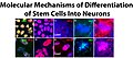 Differentiation of Stem Cells Into Neurons.jpg 1 884 × 835; 845 KB
-
 Drawing of Cell Wall.jpg 2 048 × 1 536; 637 KB
-
 Educatina 3.jpg 828 × 442; 37 KB
-
 Embolized Uterus (44507244114).jpg 1 350 × 1 124; 646 KB
-
 Endomembrane system diagram gl.svg 653 × 518; 113 KB
-
 Erythrocytes (red blood cells) Rouleaux stacking.gif 440 × 440; 3,3 MB
-
 Eukaryotic Cell (animal)-lv.jpg 1 247 × 856; 139 KB
-
 Eukaryotic cells.jpg 1 122 × 793; 98 KB
-
 Evolucion de células eucariotas.jpg 1 280 × 616; 93 KB
-
 Evoluciondecelulaseucariotas.jpg 1 280 × 616; 118 KB
-
 Fiber ends Cx50-1.jpg 3 432 × 3 840; 1,47 MB
-
 Fibroblastos 100x a.jpg 2 048 × 1 536; 257 KB
-
 FISH imaging of myc RNA in RPE.pdf 987 × 987; 25 KB
-
 Flagellum-beating-lv.svg 960 × 720; 30 KB
-
 Flower petal cells image 2.jpg 3 072 × 4 096; 5,6 MB
-
 Flower petal cells.jpg 3 072 × 4 096; 4,75 MB
-
 Fractals in cells of a flower.jpg 640 × 480; 84 KB
-
 Gas vesicle TEM.pdf 487 × 562; 659 KB
-
 Gas vesicles.pdf 600 × 881; 611 KB
-
 Giemsa Stain Macrophage Illustration.png 2 082 × 1 807; 2,9 MB
-
 Glomerulus Diameter Measurement.jpg 501 × 168; 24 KB
-
 Grapes.png 1 024 × 1 024; 662 KB
-
 Halobacterial Gas Vesicles.pdf 487 × 779; 758 KB
-
 HEK cell transfection.jpg 680 × 512; 184 KB
-
 HEK293 cells.png 500 × 387; 217 KB
-
 HEK293FTinvitro.jpg 311 × 313; 29 KB
-
 HeLa Cell (5940515305).jpg 800 × 484; 175 KB


_(20183823538).jpg)
_(20184969169).jpg)


.jpg)


_(20207617933).jpg)
_(20819051202).jpg)


.jpg)


_(14592593079).jpg)


.jpg)

_Rouleaux_stacking.gif)
-lv.jpg)


.jpg)
.jpg)