Category:Embryology
跳转到导航
跳转到搜索
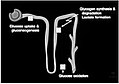
branch of biology studying prenatal biology .png) | |||||
| 上传媒体 | |||||
| 隶属于 | |||||
|---|---|---|---|---|---|
| 上级分类 | |||||
| 所属实体 | |||||
| |||||
子分类
本分类有以下58个子分类,共有58个子分类。
.
A
- Archenteron (37 F)
B
- Blastocoel (19 F)
- Branchial arches (3 F)
- Branchial region (12 F)
C
- Connecting stalk (14 F)
E
- Embryo loss (14 F)
- Embryonic induction (16 F)
- Embryonic lethality (9 F)
- Embryons desséchés (1 P, 9 F)
- Epiblast (40 F)
F
G
- Germ disc (1 F)
- Gubernaculum testis (1 F)
H
- Hypoblast (38 F)
I
- Intermediate trophoblast (8 F)
- Invagination (8 F)
K
- Koller's sickle (13 F)
M
- Mesoblast (12 F)
N
- Neural groove (28 F)
- Nidation (6 F)
O
P
- Periblast (10 F)
- Pharyngeal arches (5 F)
- Preformation theory (4 F)
- Primitive groove (3 F)
- Primitive node (21 F)
- Primitive streak (100 F)
R
- Rathke's pouch (1 F)
S
- Standard Event System (110 F)
- Stomodeum (7 F)
T
U
V
- Vitelline duct (10 F)
W
- Wolffian duct (3 F)
分类“Embryology”中的媒体文件
以下200个文件属于本分类,共473个文件。
(上一页)(下一页)-
 'The development of the blood vessels of the chick' Wellcome M0016997.jpg 2,563 × 4,118;1.95 MB
-
 4 week embryo.jpg 2,093 × 1,693;910 KB
-
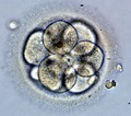 8cell embryo.tif 2,001 × 1,769;10.13 MB
-
 A lateral view 4 days post fertilisation zebrafish brain.jpg 4,263 × 2,387;1.14 MB
-

-
A-Computational-Clonal-Analysis-of-the-Developing-Mouse-Limb-Bud-pcbi.1001071.s010.ogv 18秒, 917 × 719;8.64 MB
-
 Absence of the portal system in a first trimester human.jpg 3,048 × 1,840;2.58 MB
-
 Agenesis of ductus venosus human.jpg 3,240 × 1,000;2.1 MB
-
 Amniote embryo ku.jpg 1,162 × 871;206 KB
-
 Amniote embryo.jpg 1,268 × 954;415 KB
-
 An example of hepatoblast delamination..jpg 400 × 189;54 KB
-
 Anatomic and histopathological aspects of FT organs human.jpg 3,948 × 1,292;4.99 MB
-
 Anatomische Hefte (1907) (18173584721).jpg 3,272 × 2,416;1,000 KB
-
 Anatomy and physiology of animals A reflex arc az.jpg 549 × 272;37 KB
-
 Anatomy and physiology of animals An ovum.jpg 547 × 149;14 KB
-

-

-
 Annales des Sciences Naturelles Botaniques (1850) (17787900123).jpg 2,432 × 3,732;1.48 MB
-
 Annales des Sciences Naturelles Botaniques (1850) (18220743868).jpg 2,572 × 4,244;1.75 MB
-
 Annales des Sciences Naturelles Botaniques (1850) (18408562285).jpg 2,702 × 4,489;866 KB
-
 Annales des Sciences Naturelles Botaniques (1850) (18410223821).jpg 2,618 × 4,448;776 KB
-
 Ant eggs and larvae illustration by Van Leeuwenhoek M0016636.jpg 3,030 × 3,923;3.39 MB
-
 Atv1.jpg 781 × 573;52 KB
-
 Atv12.jpg 360 × 253;20 KB
-
 Atv13.jpg 363 × 318;76 KB
-
 Atv4.jpg 687 × 566;41 KB
-
 Atv5.jpg 742 × 595;81 KB
-
 Atv6.jpg 724 × 425;57 KB
-
 Atv7.jpg 642 × 559;76 KB
-
 Atv9.jpg 352 × 527;43 KB
-
 Aves Neural tube and somites in chick embryo.jpg 1,472 × 1,144;674 KB
-
 Axial twist in zebrafish embryo.pdf 1,204 × 866;33 KB
-
 AxialTwistSchema.png 493 × 646;83 KB
-
 Beatrice Mintz (b. 1921) (6891505741).jpg 1,512 × 2,000;505 KB
-
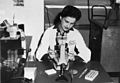 Beatrice Mintz at microscope.jpg 1,449 × 1,003;287 KB
-

-
-

-

-

-
 Blastocisto (Estructura).jpg 570 × 242;89 KB
-
 Blastocyst.JPG 500 × 375;106 KB
-
 Blastulation.png 799 × 344;100 KB
-
 Bootstrapping tableau.jpg 2,404 × 1,464;570 KB
-
 Brine shrimp cyst.jpg 146 × 145;30 KB
-
 Brockhaus-Efron Exogastrula 1.jpg 489 × 313;25 KB
-
 Brockhaus-Efron Exogastrula 2.jpg 310 × 516;50 KB
-
 Brockhaus-Efron Exoneurula 1.jpg 563 × 342;49 KB
-
 Brockhaus-Efron Exoneurula 2.jpg 436 × 630;43 KB
-
 Brockhaus-Efron Experimental Embryology 1.jpg 254 × 254;15 KB
-
 Brockhaus-Efron Experimental Embryology 10.jpg 374 × 377;61 KB
-
 Brockhaus-Efron Experimental Embryology 2.jpg 450 × 237;22 KB
-
 Brockhaus-Efron Experimental Embryology 3.jpg 415 × 404;36 KB
-
 Brockhaus-Efron Experimental Embryology 4.jpg 412 × 280;30 KB
-
 Brockhaus-Efron Experimental Embryology 5.jpg 540 × 189;29 KB
-
 Brockhaus-Efron Experimental Embryology 6.jpg 650 × 286;47 KB
-
 Brockhaus-Efron Experimental Embryology 7.jpg 665 × 588;73 KB
-
 Brockhaus-Efron Experimental Embryology 8.jpg 374 × 682;55 KB
-
 Brockhaus-Efron Experimental Embryology 9.jpg 385 × 383;58 KB
-
 C. elegans embryo development.tif 1,740 × 2,000;9.98 MB
-
 Cercetari de embriologie experimentala (1958) (19968140993).jpg 1,310 × 2,050;907 KB
-
 Chaturvedi-Simulation.jpg 960 × 720;26 KB
-
 Chemical embryology (1931) (20416409820).jpg 1,884 × 2,222;1.4 MB
-
 Chemical embryology (1931) (20416494850).jpg 1,712 × 1,340;280 KB
-
 Chemical embryology (1931) (20604356705).jpg 794 × 1,386;291 KB
-
 Conger type callus 3ms White Light.TIF 2,048 × 1,536;9.01 MB
-
 Contributions to embryology (20503173019).jpg 1,372 × 3,028;734 KB
-
 Contributions to embryology (20680611872).jpg 2,036 × 2,884;698 KB
-
 Contributions to embryology (20689887595).jpg 2,212 × 2,826;1.15 MB
-
 Contributions to embryology (20690137775).jpg 2,088 × 1,504;513 KB
-
 Contributions to embryology (20696603441).jpg 2,265 × 2,900;716 KB
-
 Contributions to embryology (20696681371).jpg 3,698 × 2,114;1.61 MB
-
 Contributions to embryology (20696849551).jpg 2,126 × 2,824;996 KB
-
 Contributions to embryology (IA contributionstoe09carn).pdf 981 × 1,229,684页;40.44 MB
-
 CpG methylation in mouse development.png 1,660 × 807;188 KB
-
 Cresta neurale.png 920 × 1,172;213 KB
-
 Cristatus implant.png 768 × 748;285 KB
-
 Critique of the Theory of Evolution Fig 007.jpg 414 × 413;37 KB
-
 Critique of the Theory of Evolution Fig 009.jpg 586 × 292;31 KB
-
 Critique of the Theory of Evolution Fig 011.jpg 460 × 208;17 KB
-
 Critique of the Theory of Evolution Fig 012.jpg 700 × 584;63 KB
-
 Critique of the Theory of Evolution Fig 048.jpg 520 × 503;45 KB
-
 Da Vinci Studies of Embryos Luc Viatour.jpg 1,443 × 2,121;2.66 MB
-
 Denticlebands.png 320 × 470;97 KB
-
 Desarrollo de la Notocorda en Embrión Humano.PNG 738 × 555;704 KB
-
 Descartes; A Treatise on the formation of the foetus Wellcome L0017414.jpg 1,201 × 1,569;900 KB
-
 Descartes; A Treatise on the formation of the foetus Wellcome L0017415.jpg 1,865 × 980;328 KB
-
 Descartes; A Treatise on the formation of the foetus Wellcome L0017416.jpg 1,254 × 1,526;532 KB
-
 Descartes; A Treatise on the formation of the foetus Wellcome L0017417.jpg 1,228 × 1,546;476 KB
-
 Descartes; A Treatise on the formation of the foetus Wellcome L0017418.jpg 1,113 × 1,659;597 KB
-
 Descartes; Quelle est la fabrique de ses nerfs. Wellcome M0014443.jpg 2,675 × 3,974;967 KB
-
 Deuterostomes.png 1,992 × 927;508 KB
-
 Deuterostomia.png 590 × 263;104 KB
-
 Developing placenta.jpg 678 × 334;81 KB
-
 Development of embryonic nephrons.png 4,059 × 3,000;1.8 MB
-
 Development of plant embryos Wellcome M0016635.jpg 4,291 × 2,739;2.68 MB
-
 Diagram showing human embryo grades for in vitro fertilisation (IVF).jpg 1,699 × 1,139;212 KB
-
 Diaphragma-embryo.png 596 × 533;91 KB
-

-

-
 Differentiation Tree of the Axolotl.jpg 1,254 × 969;372 KB
-
 Dorsal lip transplantation in a salamander embryo.png 728 × 838;649 KB
-
 Déterminantscyto.png 587 × 394;295 KB
-
 E Filogenia metazoa celoma.png 982 × 818;122 KB
-
 Early embryonic development.JPG 807 × 860;179 KB
-
 Early stages in the development of the sheep embryo. Wellcome M0011382.jpg 2,528 × 4,170;2.12 MB
-
 Ecografía 4D - Feto 14semanas C.jpg 584 × 399;34 KB
-
 EHR-BBII.jpg 1,500 × 1,125;304 KB
-
 Elefetusus.jpg 1,910 × 1,352;276 KB
-
 Embrião de Rã - Secção da Cabeça.png 776 × 520;838 KB
-
 Embrião de Rã - Secção do Abdómen.png 639 × 805;1.12 MB
-
 Embrião de Rã - Secção do Tórax.png 560 × 790;767 KB
-
 Embryo after first 24 hours. Wellcome M0011396.jpg 2,461 × 4,403;1.76 MB
-
 Embryo after first 24 hours. Wellcome M0011397.jpg 4,801 × 2,256;812 KB
-
 Embryo after first 24 hours. Wellcome M0011398.jpg 5,870 × 1,830;2.06 MB
-
 Embryo at three months. Brain and spinal cord exposed. Wellcome M0011400.jpg 2,078 × 5,248;1.34 MB
-
 Embryo developing2.png 458 × 258;88 KB
-

-
 Embryo in flower.png 3,000 × 3,006;2.97 MB
-
 Embryo of a chick Wellcome M0010698.jpg 3,562 × 3,140;2.02 MB
-
 Embryo, showing development of central nervous system Wellcome M0011399.jpg 2,148 × 5,308;2.04 MB
-
 Embryological development of the human venous system.png 2,980 × 1,672;1.37 MB
-
 Embryologie Zwerchfell.png 2,871 × 1,472;682 KB
-
 Embryology (1949) (20662846714).jpg 1,620 × 1,944;647 KB
-
 Embryology (1949) (20664399993).jpg 1,800 × 2,104;398 KB
-
 Embryology (1949) (21097805768).jpg 1,832 × 1,264;878 KB
-
 Embryology (1949) (21285445665).jpg 1,344 × 2,588;348 KB
-
 Embryology (1949) (21285693065).jpg 1,035 × 1,115;581 KB
-
Embryology 3d.ogv 11分25秒, 1,280 × 720;87.12 MB
-
 Embryology and Cytology Drawings 1919-1920 Wellcome L0024688.jpg 1,872 × 1,114;883 KB
-
 Embryology and Cytology Drawings Wellcome L0024689.jpg 1,874 × 1,093;851 KB
-
 Embryology-childs-depiction.png 1,280 × 983;589 KB
-
 Embryology; Theoria Generationis Wellcome M0011665.jpg 2,965 × 3,616;4.96 MB
-
 EmbryonBlastocyste.jpg 1,489 × 1,302;194 KB
-
 EmbryonDisqueEmbryonnaire.jpg 1,576 × 1,378;278 KB
-
 EmbryonGastrulation.jpg 1,556 × 1,086;317 KB
-
 EmbryonGastrulationII.jpg 1,432 × 1,273;361 KB
-
 EmbryonGastrulationIII.jpg 1,585 × 1,599;474 KB
-
 EmbryonGastrulationIV.jpg 1,661 × 1,321;427 KB
-
 Embryonic Development CNS (ja).png 700 × 650;186 KB
-
 Embryonic Development CNS ar.png 350 × 325;446 KB
-
 Embryonic Development CNS.png 350 × 325;23 KB
-
 Embryonic dinosaur bones.tif 2,344 × 1,774;15.86 MB
-
 Embryonic spinal cord.jpg 457 × 277;21 KB
-
 EmbryonVitellinPrimaire.jpg 1,676 × 2,025;735 KB
-
 EmbryonVitellinsecondaire.jpg 1,685 × 2,120;789 KB
-
 EmbryoScope.jpg 6,544 × 4,367;1.98 MB
-
 End of week 4 Embryo with somites nltxt.jpg 925 × 674;203 KB
-
 End of week 4 Embryo with somites.jpg 960 × 720;121 KB
-
 Endoderm2 hr.png 400 × 290;62 KB
-
 Endodermaglandularumperiarteriosarum.png 670 × 228;150 KB
-
 Engraving; Growth of chick embryo at days 19-20, 1625. Wellcome L0007939.jpg 1,100 × 1,712;780 KB
-
 Engraving; Growth of chick embryo at days 21-24, 1625. Wellcome L0007940.jpg 1,050 × 1,689;730 KB
-
 Ernst Haeckel, Anthropogenie. Wellcome L0027291.jpg 1,102 × 1,716;816 KB
-
 Ernst Haeckel, Anthropogenie. Wellcome L0027292.jpg 1,360 × 2,070;1.14 MB
-
 Evolution du follicule dans l'ovaire.jpg 1,086 × 655;108 KB
-
 Evolution of the avian ankle.jpg 1,773 × 1,512;275 KB
-
 F. Ruysch "Thesaurus..."; botanical preparations Wellcome M0016627.jpg 2,913 × 3,939;4.12 MB
-
 F. Ruysch "Thesaurus..."; botanical preparations Wellcome M0016631.jpg 2,498 × 4,443;3.46 MB
-
 Fertilisation.gif 600 × 421;16 KB
-
 Fertilisation2.png 535 × 983;442 KB
-
 Fertilized egg.png 500 × 375;87 KB
-
 Fetal circulation.jpg 3,600 × 3,591;7.13 MB
-
 FetalMembranes1L.jpg 424 × 400;57 KB
-
 Figure 27 02 05.jpg 1,117 × 738;593 KB
-
 Filogenia dos Metazoa e Cavidade Celômica.png 982 × 818;122 KB
-
 Filogenia dos Metazoa e celoma .png 818 × 982;159 KB
-
 Filogenia dos metazoa.jpg 1,056 × 816;114 KB
-
 Filogenia Metazoa .png 982 × 809;90 KB
-
 FPimage.jpg 960 × 720;43 KB
-
 Frog's egg, immature fish and blood corpuscles, etc. Wellcome M0016632.jpg 4,800 × 2,221;2.84 MB
-
 Gad1 transcripts in the developing vibrissae.jpg 1,200 × 2,018;131 KB
-
Gastrulation in 3D.ogv 6分19秒, 1,280 × 720;22.73 MB
-
 Gastrulation.png 801 × 486;63 KB
-
 Gastrulação.png 695 × 349;13 KB
-
 Gema1.png 984 × 143;74 KB
-
 Genitalia embryonis feminei.png 494 × 480;59 KB
-
 GI Trace Development.gif 1,018 × 768;38.73 MB
-
 Gonadotrofas Capilar.png 1,016 × 1,026;1.7 MB
-
 Gonadotrofas FSH LH.png 686 × 336;274 KB
-
 Gonadotropas Capilar.png 584 × 602;316 KB
-
 Gonadotropas Stellates.jpg 776 × 628;245 KB
-
 GR&PGCs.png 1,377 × 688;187 KB
-
 Gray1106.png 730 × 500;45 KB
-
 Gray1112.png 526 × 500;35 KB
-
 Gray12.png 450 × 181;12 KB
-
 Gray13.png 215 × 245;25 KB
-
 Gray13a.jpg 215 × 245;31 KB
-
 Gray15.png 400 × 485;79 KB
-
 Gray18.png 379 × 652;62 KB
-
 Gray29.png 500 × 306;15 KB
-
 Gray36-ar.png 450 × 327;65 KB
-
 Gray36.png 450 × 320;25 KB
-
 Gray39 cs.png 576 × 384;50 KB
-
 Gray39 sp.PNG 500 × 384;38 KB
-
 Gray39.png 500 × 384;41 KB
-
 Gray56.png 567 × 442;20 KB
-
 Gray65 colour.jpg 550 × 316;139 KB
-
 Gray65.png 550 × 316;29 KB
-
 Gray69.png 550 × 304;22 KB


_(18173584721).jpg)


_(17787900123).jpg)
_(18220743868).jpg)
_(18408562285).jpg)
_(18410223821).jpg)


_(6891505741).jpg)

_(19743465713).jpg)
_(20177724519).jpg)

_(1893)_(20354053046).jpg)
_(1899)_(20380332505).jpg)
.jpg)


_(19968140993).jpg)

_(20416409820).jpg)
_(20416494850).jpg)
_(20604356705).jpg)
.jpg)
.jpg)
.jpg)
.jpg)
.jpg)
.jpg)
.jpg)


.jpg)

_165_174_Entwicklung_der_menschlichen_Frucht_bis_zum_Beginn_des_dritten_Monats.png)


_after_72_hours_of_incubation.jpg)


_(20662846714).jpg)
_(20664399993).jpg)
_(21097805768).jpg)
_(21285445665).jpg)
_(21285693065).jpg)


.png)


