Category:Images from Cancer Research UK
Jump to navigation
Jump to search
These images have been released as part of an open knowledge project by Cancer Research UK. If re-used, attribute to Cancer Research UK / Wikimedia Commons. Please do not add files to or remove them from this category, except for translated versions etc. Check total, 4 August 2014, 392 files (2 duplicates)
Subcategories
This category has the following 6 subcategories, out of 6 total.
A
D
I
P
Media in category "Images from Cancer Research UK"
The following 200 files are in this category, out of 683 total.
(previous page) (next page)-
10 Things You Didn't Know About Pancreatic Cancer.webm 1 min 56 s, 1,920 × 1,080; 8.14 MB
-
 13.04.14CRUK 0060(2).jpg 6,000 × 4,000; 6.76 MB
-
 13.04.14CRUK 0466(1).jpg 6,000 × 4,000; 7.77 MB
-
 13.04.14CRUK 0564(1).jpg 6,000 × 4,000; 8.24 MB
-
 Ainslee-clarke-copy.jpg 1,322 × 2,000; 914 KB
-
 Alcohol causes 7 types of cancer.svg 610 × 688; 124 KB
-
 BCPcrukBristolopening027x(1).jpg 4,288 × 2,848; 1.58 MB
-
 Bill-house-copy (1).jpg 1,323 × 2,000; 1.9 MB
-
 Bill-house.jpg 1,407 × 2,000; 1,020 KB
-
 Breast cancer incidence by anatomical site (females)-ar.png 650 × 587; 72 KB
-
 Breast cancer incidence by anatomical site (females).svg 650 × 587; 17 KB
-
 Bunido-pontecorno.jpg 1,940 × 2,409; 531 KB
-
 Cambridge 84 (Print Quality Version(Large)) (crctprod 005443 Revision-1)(1).jpg 2,100 × 1,370; 805 KB
-
 Cambridge 96 (Print Quality Version(Large)) (crctprod 005448 Revision-1)(1).jpg 2,100 × 1,432; 912 KB
-
 Clare-hall.jpg 2,800 × 1,825; 1.96 MB
-
 CR 03-11 0256 (Print Quality Version(Large)) (crctprod 009830 Revision-1)(1).jpg 2,100 × 1,400; 495 KB
-
 Crowd (wave) - L(1).jpg 1,280 × 857; 1.24 MB
-
 CRUK-25996 1(1).tif 5,760 × 3,840, 2 pages; 63.3 MB
-
 CRUK-26028(1).tif 5,760 × 3,840, 2 pages; 63.3 MB
-
 CRUK-26047(1).tif 5,760 × 3,840, 2 pages; 63.3 MB
-
 CRUK-26233(1).tif 5,760 × 3,840, 2 pages; 63.31 MB
-
 CRUK-test-25959(1).tif 5,632 × 3,755, 2 pages; 60.53 MB
-
-

-
 Diagram 1 of 2 showing stage 2A breast cancer CRUK 003-ar.png 375 × 314; 36 KB
-
 Diagram 1 of 2 showing stage 2A breast cancer CRUK 003.svg 375 × 314; 44 KB
-
 Diagram 1 of 2 showing stage 3A vulval cancer CRUK 496.svg 425 × 230; 56 KB
-
 Diagram 1 of 2 showing stage 3B breast cancer CRUK 004-ar.png 375 × 383; 42 KB
-
 Diagram 1 of 2 showing stage 3B breast cancer CRUK 004.svg 375 × 383; 23 KB
-
 Diagram 1 of 2 showing stage 3B lung cancer CRUK 005.svg 425 × 345; 541 KB
-
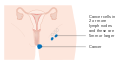 Diagram 1 of 2 showing stage 3B vulval cancer CRUK 498.svg 425 × 230; 56 KB
-
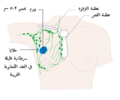 Diagram 1 of 3 showing stage 2B breast cancer CRUK 006-ar.png 375 × 314; 34 KB
-
 Diagram 1 of 3 showing stage 2B breast cancer CRUK 006.svg 375 × 314; 42 KB
-
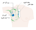 Diagram 1 of 3 showing stage 3A breast cancer CRUK 007-ar.png 375 × 310; 32 KB
-
 Diagram 1 of 3 showing stage 3A breast cancer CRUK 007.svg 375 × 310; 43 KB
-
 Diagram 1 of 3 showing stage 3A lung cancer CRUK 008.svg 425 × 284; 492 KB
-
 Diagram 1 of 3 showing stage 3A oesophageal cancer CRUK 479.svg 375 × 257; 533 KB
-
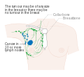 Diagram 1 of 3 showing stage 3C breast cancer CRUK 399.svg 375 × 348; 43 KB
-
 Diagram 1 of 3 showing stage 3C oesophageal cancer CRUK 481.svg 375 × 270; 535 KB
-
 Diagram 1 of skinning vulvectomy CRUK 469.svg 500 × 312; 59 KB
-
 Diagram 2 of 2 showing stage 2A breast cancer CRUK 009-ar.png 368 × 259; 33 KB
-
 Diagram 2 of 2 showing stage 2A breast cancer CRUK 009.svg 368 × 259; 38 KB
-
 Diagram 2 of 2 showing stage 3A vulval cancer CRUK 497.svg 425 × 230; 56 KB
-
 Diagram 2 of 2 showing stage 3B breast cancer CRUK 010-ar.png 375 × 313; 30 KB
-
 Diagram 2 of 2 showing stage 3B breast cancer CRUK 010.svg 375 × 313; 49 KB
-
 Diagram 2 of 2 showing stage 3B lung cancer CRUK 011.svg 425 × 340; 543 KB
-
 Diagram 2 of 2 showing stage 3B vulval cancer CRUK 499.svg 425 × 230; 56 KB
-
 Diagram 2 of 3 showing stage 2B breast cancer CRUK 012-ar.png 375 × 306; 34 KB
-
 Diagram 2 of 3 showing stage 2B breast cancer CRUK 012.svg 375 × 306; 51 KB
-
 Diagram 2 of 3 showing stage 3A breast cancer CRUK 013-ar.png 375 × 310; 34 KB
-
 Diagram 2 of 3 showing stage 3A breast cancer CRUK 013.svg 375 × 310; 44 KB
-
 Diagram 2 of 3 showing stage 3A lung cancer CRUK 014.svg 425 × 311; 493 KB
-
 Diagram 2 of 3 showing stage 3A oesophageal cancer CRUK 482.svg 375 × 235; 38 KB
-
 Diagram 2 of 3 showing stage 3C breast cancer CRUK 400.svg 375 × 349; 51 KB
-
 Diagram 2 of 3 showing stage 3C oesophageal cancer CRUK 483.svg 375 × 267; 608 KB
-
 Diagram 2 of skinning vulvectomy CRUK 470-ar.png 400 × 301; 38 KB
-
 Diagram 2 of skinning vulvectomy CRUK 470.svg 400 × 301; 19 KB
-
 Diagram 3 of 3 showing stage 2B breast cancer CRUK 015-ar.png 375 × 314; 33 KB
-
 Diagram 3 of 3 showing stage 2B breast cancer CRUK 015.svg 375 × 314; 52 KB
-
 Diagram 3 of 3 showing stage 3A breast cancer CRUK 016-ar.png 375 × 310; 35 KB
-
 Diagram 3 of 3 showing stage 3A breast cancer CRUK 016.svg 375 × 310; 51 KB
-
 Diagram 3 of 3 showing stage 3A lung cancer CRUK 017.svg 425 × 339; 541 KB
-
 Diagram 3 of 3 showing stage 3A oesophageal cancer CRUK 484.svg 375 × 235; 38 KB
-
 Diagram 3 of 3 showing stage 3C breast cancer CRUK 401.svg 375 × 347; 44 KB
-
 Diagram 3 of 3 showing stage 3C oesophageal cancer CRUK 485.svg 375 × 264; 31 KB
-
 Diagram 3 of skinning vulvectomy CRUK 471.svg 400 × 301; 18 KB
-
 Diagram of a 3 in 1 incision vulvectomy CRUK 018-ar.svg 500 × 312; 159 KB
-
 Diagram of a 3 in 1 incision vulvectomy CRUK 018.svg 500 × 312; 19 KB
-
 Diagram of a breast implant CRUK 402.svg 375 × 305; 15 KB
-
 Diagram of a chromosome in a cell CRUK 019.svg 334 × 328; 10 KB
-
 Diagram of a gene on a chromosome CRUK 020.svg 345 × 322; 9 KB
-
 Diagram of a larkel CRUK 021.svg 375 × 249; 84 KB
-
 Diagram of a lymph node CRUK 022 ku.svg 375 × 306; 213 KB
-
 Diagram of a lymph node CRUK 022.svg 375 × 306; 176 KB
-
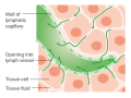 Diagram of a lymphatic capillary CRUK 023.svg 375 × 280; 78 KB
-
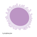 Diagram of a lymphocyte CRUK 024.svg 283 × 279; 13 KB
-
-

-

-
-
 Diagram of a plasma cell CRUK 409.svg 344 × 318; 3 KB
-
 Diagram of a platelet CRUK 407.svg 322 × 307; 2 KB
-
 Diagram of a red blood cell CRUK 467.svg 286 × 288; 69 KB
-
 Diagram of a transitional cell CRUK 027.svg 375 × 272; 611 KB
-
 Diagram of a white blood cell CRUK 028.svg 389 × 384; 695 KB
-
 Diagram of an astrocyte - a type of glial cell CRUK 029.svg 344 × 301; 43 KB
-
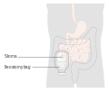 Diagram of an ileostomy with a bag CRUK 030.svg 375 × 320; 45 KB
-
 Diagram of an inflatable breast implant cruk 403.svg 375 × 308; 24 KB
-
 Diagram of an osteocyte - a type of bone cell CRUK 031.svg 375 × 300; 459 KB
-
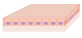 Diagram of basal cells CRUK 410.svg 375 × 176; 420 KB
-
 Diagram of bone marrow CRUK 462.svg 375 × 444; 507 KB
-
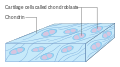 Diagram of cartilage cells called chondroblasts CRUK 032.svg 375 × 209; 24 KB
-
 Diagram of epithelial cells CRUK 033.svg 375 × 197; 530 KB
-
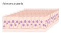 Diagram of glandular cells CRUK 034.svg 375 × 215; 2.17 MB
-
 Diagram of muscle cells CRUK 035.svg 375 × 204; 970 KB
-
 Diagram of squamous cells CRUK 036.svg 375 × 160; 686 KB
-
 DIagram of the different types of soft tissue in the body CRUK 037.svg 375 × 411; 457 KB
-
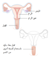
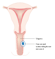
 Diagram of the female human pelvis cross section.svg 266 × 334; 23 KB
-
 Diagram of the gastro oesophageal junction CRUK 038.svg 375 × 338; 20 KB
-
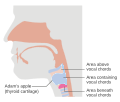 Diagram of the larynx CRUK 039.svg 384 × 325; 85 KB
-
 Diagram of the lung showing pleural mesothelioma CRUK 458.svg 375 × 360; 68 KB
-
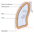 Diagram of the lung showing the pleura CRUK 459.svg 375 × 371; 60 KB
-
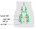 Diagram of the lymph nodes in the pelvis CRUK 040-ar.png 375 × 310; 55 KB
-
 Diagram of the lymph nodes in the pelvis CRUK 040.svg 375 × 310; 93 KB
-
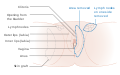
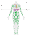 Diagram of the lymphatic system CRUK 041.svg 375 × 429; 318 KB
-
 Diagram of the male urinary system CRUK 042.svg 375 × 350; 18 KB
-

-
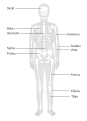 Diagram of the skeleton CRUK 044.svg 375 × 518; 79 KB
-
 Diagram of the small bowel 01 CRUK 045.svg 375 × 304; 38 KB
-
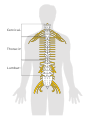 Diagram of the spinal cord CRUK 046.svg 402 × 542; 164 KB
-

-

-
 Diagram of the testicles CRUK 048.svg 375 × 270; 174 KB
-
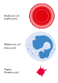 Diagram of three different types of blood cell CRUK 049.svg 347 × 493; 723 KB
-
 Diagram of what is in blood CRUK 050.svg 250 × 350; 4 KB
-
 Diagram showing a bone marrow biopsy CRUK 051.svg 375 × 318; 170 KB
-
 Diagram showing a brain shunt CRUK 052.svg 375 × 365; 81 KB
-
 Diagram showing a bronchoscopy CRUK 053.svg 375 × 383; 228 KB
-

-
 Diagram showing a burr hole biopsy CRUK 055.svg 375 × 387; 80 KB
-
-

-
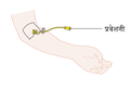 Diagram showing a cannula CRUK 058-hi.png 360 × 247; 14 KB
-
 Diagram showing a cannula CRUK 058-hi.svg 360 × 247; 22 KB
-
 Diagram showing a cannula CRUK 058-multilingual1.svg 360 × 247; 19 KB
-
 Diagram showing a cannula CRUK 058-pa.png 360 × 247; 14 KB
-
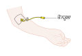 Diagram showing a cannula CRUK 058-pa.svg 360 × 247; 22 KB
-
 Diagram showing a cannula CRUK 058-ta.png 360 × 247; 14 KB
-
 Diagram showing a cannula CRUK 058.png 360 × 247; 13 KB
-
 Diagram showing a cannula CRUK 058.svg 360 × 247; 19 KB
-
 Diagram showing a central line CRUK 059.svg 375 × 250; 42 KB
-
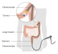
 Diagram showing a colonoscopy CRUK 060.svg 375 × 282; 84 KB
-
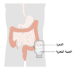 Diagram showing a colostomy with a bag CRUK 061-ar.png 1,200 × 1,024; 122 KB
-
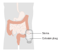 Diagram showing a colostomy with a bag CRUK 061.svg 375 × 320; 56 KB
-
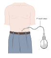
 Diagram showing a continent urinary diversion CRUK 062.svg 375 × 360; 60 KB
-
 Diagram showing a craniotomy CRUK 063.svg 376 × 393; 1.42 MB
-
 Diagram showing a cystoscopy for a man and a woman CRUK 064-ar.png 375 × 555; 72 KB
-
 Diagram showing a cystoscopy for a man and a woman CRUK 064.svg 375 × 555; 58 KB
-
 Diagram showing a double helix of a chromosome CRUK 065-hi.png 301 × 388; 46 KB
-
 Diagram showing a double helix of a chromosome CRUK 065-hi.svg 300 × 387; 19 KB
-
 Diagram showing a double helix of a chromosome CRUK 065.svg 300 × 387; 15 KB
-
-
 Diagram showing a lobectomy of the thyroid gland CRUK 067.svg 360 × 439; 276 KB
-
-
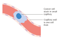
 Diagram showing a malignant tumour CRUK 069.svg 375 × 305; 212 KB
-
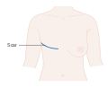 Diagram showing a mastectomy scar CRUK 447.svg 332 × 272; 11 KB
-

-
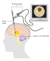 Diagram showing a neuroendoscopy CRUK 475.svg 375 × 451; 313 KB
-
 Diagram showing a person on a ventilator CRUK 460.svg 375 × 325; 34 KB
-
 Diagram showing a PICC line CRUK 071.svg 375 × 266; 57 KB
-
-

-
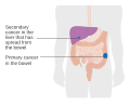 Diagram showing a primary and secondary cancer CRUK 074.svg 375 × 301; 37 KB
-
 Diagram showing a prostate biopsy CRUK 472.svg 375 × 370; 471 KB
-

-

-
-

-
 Diagram showing a rectosigmoid pouch CRUK 077.svg 375 × 365; 98 KB
-
-

-

-

-
-
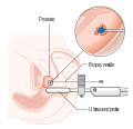 Diagram showing a transperineal prostate biopsy CRUK 473.svg 380 × 368; 323 KB
-
 Diagram showing a tumour causing spinal cord compression CRUK 081.svg 309 × 251; 912 KB
-

-
 Diagram showing a urinary catheter in a man CRUK 084.svg 375 × 435; 48 KB
-

-
 Diagram showing a urinary catheter in a woman CRUK 085.svg 375 × 435; 28 KB
-
 Diagram showing a voice valve CRUK 086.svg 375 × 350; 24 KB
-

-
-
 Diagram showing a wide local excision of the vulva CRUK 088.svg 500 × 312; 18 KB
-
 Diagram showing a woman having a mammogram CRUK 089.svg 375 × 314; 48 KB
-
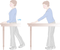
 Diagram showing abdominal breathing CRUK 090.svg 286 × 595; 114 KB
-
 Diagram showing abdominoperineal resection of the anus CRUK 091.svg 375 × 330; 50 KB
-
 Diagram showing advanced bladder cancer CRUK 441.svg 375 × 448; 86 KB
-

-

-
 Diagram showing an above knee amputation CRUK 094.svg 375 × 285; 307 KB
-
 Diagram showing an airway stent CRUK 095.svg 375 × 367; 521 KB
-
 Diagram showing an antibody CRUK 096.svg 270 × 300; 3 KB
-
-
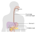
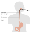 Diagram showing an endoscopy CRUK 098.svg 375 × 410; 17 KB
-

-
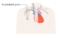 Diagram showing an implantable port CRUK 101.svg 375 × 242; 50 KB
-
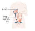
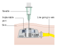 Diagram showing an implantable port under the skin CRUK 100.svg 375 × 314; 11 KB
-
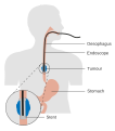 Diagram showing an oesophageal stent being put in CRUK 495.svg 375 × 425; 189 KB
-
 Diagram showing an oesophageal stent CRUK 491.svg 375 × 425; 189 KB
-
 Diagram showing before and after a partial nephrectomy CRUK 102.svg 375 × 625; 104 KB
-

-

-
 Diagram showing before and after a radical nephrectomy CRUK 104.svg 375 × 627; 252 KB
-

-
 Diagram showing before and after a total oesophagectomy CRUK 105.svg 375 × 449; 19 KB
-
 Diagram showing before and after a total thyroidectomy CRUK 106.svg 358 × 436; 348 KB
-
-
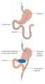 Diagram showing before and after stomach bypass surgery CRUK 108.svg 375 × 693; 20 KB
-

.jpg)
.jpg)
.jpg)


.jpg)
.jpg)

-ar.png)
.svg)

)_(crctprod_005443_Revision-1)(1).jpg)
)_(crctprod_005448_Revision-1)(1).jpg)

)_(crctprod_009830_Revision-1)(1).jpg)
_-_L(1).jpg)
_CRUK_001.svg)


_CRUK_054.svg)

_CRUK_056.svg)


_CRUK_097.svg)

_CRUK_099.svg)


