Category:Immunology
跳转到导航
跳转到搜索
医学分支 _(14788924923).jpg) | |||||
| 上传媒体 | |||||
| 隶属于 | |||||
|---|---|---|---|---|---|
| 上级分类 | |||||
| 可分为 |
| ||||
| |||||

子分类
本分类有以下50个子分类,共有50个子分类。
*
?
A
- Allograft (3 F)
- Allorecognition (3 F)
B
C
- Cellular immunity (8 F)
D
H
- Host antiviral responses (9 F)
- Human leukocyte antigen (9 F)
- Hybridoma technology (3 F)
I
- Immune evasion (24 F)
- Immune privilege (3 F)
- Immune suppression (8 F)
- Immunization (6 F)
- Immunologic adjuvants (7 F)
- Immunologic cytotoxicity (41 F)
- Immunologic factors (6 F)
- Immunological models (20 F)
- Immunological synapses (59 F)
- Immunology Wiki (2 F)
L
M
- Media from BMC Immunology (18 F)
N
- Neuroimmunology (5 F)
O
P
- Psychoneuroimmunology (1 F)
S
- SCID mice (34 F)
T
V
- Viral antigens (9 F)
分类“Immunology”中的媒体文件
以下200个文件属于本分类,共343个文件。
(上一页)(下一页)-
 "Side-chain diagram; immune bodies ..." Ehrlich Wellcome M0013394.jpg 4,997 × 2,139;1.58 MB
-
 2WINSurfaceRendering.png 640 × 515;338 KB
-
 A short treatise on anti-typhoid inoculation.djvu 2,124 × 3,374,94页;2.68 MB
-
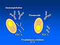 Abb2 ImmProd.jpg 720 × 540;31 KB
-
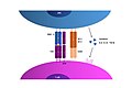
 Abb3 Nephron.jpg 720 × 540;36 KB
-
 Account of inoculations for smallpox, 1724. Wellcome M0011151.jpg 2,457 × 4,290;2 MB
-
 Activación de un linfocito T.jpg 2,160 × 1,440;148 KB
-
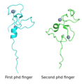 Aire protein (first- and second phd fingers).png 598 × 613;71 KB
-
 Alloreactive Antisera.PNG 312 × 399;12 KB
-
 ANA MIDBODY.jpg 1,177 × 1,026;1.47 MB
-
 ANA NEGATIVE LIVER.jpg 1,240 × 864;1.33 MB
-
 ANA NUCLEAR DOT AND AMA.jpg 1,323 × 1,026;2.16 MB
-
 ANA NUCLEAR DOTS AND NUCLEOLAR.jpg 1,330 × 893;1.51 MB
-
 ANA NUCLEOLAR 2.jpg 1,210 × 964;964 KB
-
 ANA NUCLEOLAR 3.jpg 1,130 × 1,100;1.12 MB
-
 ANA NUCLEOLAR AND MEMBRANE.jpg 1,350 × 1,131;1.69 MB
-
 ANA NUCLEOLAR LIVER.jpg 1,242 × 1,029;1.86 MB
-
 ANA NUCLEOLAR.jpg 1,231 × 974;1.42 MB
-
 ANA SPECKLED LIVER.jpg 1,435 × 969;1.54 MB
-
 ANA SPINDEL FIBER.jpg 1,018 × 1,038;1.11 MB
-
 ANAPHASE.jpg 1,131 × 756;1.08 MB
-
 Anti-Allergy Immunotherapy (hy).png 2,048 × 1,525;1 MB
-
 Anti-Allergy Immunotherapy.jpg 2,083 × 1,552;1.18 MB
-
 Antibody dependent enhancement.tif 816 × 720;153 KB
-
 ANTIBODY NEGATIVE.jpg 1,888 × 1,439;2.45 MB
-
 AntibodyX.JPG 492 × 362;20 KB
-
 Anticuerpos Monoclonales y Policlonales.png 612 × 340;20 KB
-
 Antigenerkennung.png 639 × 525;11 KB
-
 Apoptosome surface rendering.png 640 × 516;420 KB
-
 B cell central tolerance.png 838 × 578;61 KB
-
 B-raku aktivatsioon 2.png 390 × 599;66 KB
-
 B-raku aktivatsioon.png 390 × 599;66 KB
-
 B7 family ligands and CD28 family receptors.JPG 664 × 609;55 KB
-
 BASIC RIG-I STRUCTURE.png 1,687 × 949;175 KB
-
 Benefits, limitations, examples of different types of vaccines.png 1,294 × 1,506;353 KB
-
 Bioteknik.png 553 × 290;7 KB
-
 Blastocyst immunosurgery.png 1,561 × 4,000;1.18 MB
-
 Blausen 0624 Lymphocyte B cell eesti.png 600 × 600;235 KB
-
 C2orf72 Orthologs List.png 807 × 616;305 KB
-
 C3b.Opsonization.png 1,396 × 1,326;1.53 MB
-
 CauseInfiammazioni.png 546 × 590;330 KB
-
 CCR7 receptor.png 1,800 × 1,788;692 KB
-
 Cells-04-00178-g001.png 3,464 × 4,190;147 KB
-
 Cellular mechanisms of MAVS pathway.pdf 975 × 887;122 KB
-
 Centre germinatif.png 800 × 600;109 KB
-
 Centrifugadora d'immunohematologia.JPG 4,000 × 6,016;5.64 MB
-
 CENTROMERE.jpg 1,161 × 968;1.06 MB
-
 Changes in various immune cell subsets during immunosenescence.jpg 796 × 739;109 KB
-
 Chtx-Deu4.png 960 × 720;35 KB
-
 Class 1.jpg 720 × 540;99 KB
-
 CLIP Binding to MHC II.jpg 2,048 × 1,536;145 KB
-
 Clonal Deletion.png 1,440 × 816;155 KB
-
 Cluster of Differentiation mod.png 2,000 × 2,688;245 KB
-
 Clínica de inmunodeficiencias primarias.jpg 4,320 × 3,240;908 KB
-
 Commensals vs pathogens mechanisms.png 3,863 × 3,558;1.35 MB
-
 Comparison-of-Toll-pathways.png 814 × 581;127 KB
-
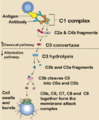 Complement pathway.png 688 × 834;89 KB
-
 Constituents of diptheria toxin, Ehrlich Wellcome M0013395.jpg 2,729 × 3,977;1.92 MB
-
 CRITHIDIA 2.jpg 1,011 × 816;768 KB
-
 Cross priming des cd8.jpg 960 × 720;36 KB
-
 Cross-presentación.png 724 × 723;191 KB
-
 Cross-presentation.png 1,008 × 750;151 KB
-
 CTL-CTLA4.png 720 × 540;7 KB
-
 Células B a lo largo de los años.jpg 4,134 × 1,772;1.38 MB
-
 Dany tisular 1.png 822 × 624;102 KB
-
 Dany tisular 2.png 806 × 636;157 KB
-
 DC-CD8.png 1,274 × 921;55 KB
-
De-Immunologie.ogg 2.2秒;21 KB
-
-

-
 Dendritic cell activation following vaccination 01.tif 4,267 × 4,267;3.93 MB
-
 Dendritic cell activation within the draining lymph node following vaccination 01.tif 4,267 × 4,267;4.74 MB
-
 Dendritic cell activation within the draining lymph node following vaccination 02.tif 4,267 × 4,267;4.83 MB
-
 Dendritic cell activation within the draining lymph node following vaccination 03.tif 4,267 × 4,267;8.64 MB
-
 Dendritic cell activation within the draining lymph node following vaccination 04.tif 4,267 × 4,267;8.58 MB
-
 Dendritic cell migration following vaccination.tif 4,267 × 4,267;3.81 MB
-
 Dendritic cell taking information from the outside.jpg 1,080 × 1,080;171 KB
-
 Dengue IgM and IgG Tests-Negative.jpg 2,340 × 4,160;2.43 MB
-
 Dien bien nhanh.JPG 478 × 709;64 KB
-
 Differenzierungsantigene und Lymphozytenreifung.jpg 1,642 × 2,306;426 KB
-
 DifImmunPatog.png 800 × 627;1.13 MB
-
 DirectELISAdiagram-page-001.JPG 1,500 × 1,125;190 KB
-
 Distribución de inmunodeficiencias primarias por tipo..jpg 507 × 448;39 KB
-
 Draining lymph node 01.tif 4,267 × 4,267;3.24 MB
-
 Droga alternatywna.png 759 × 350;66 KB
-
 Droga klasyczna.png 725 × 405;74 KB
-
 DSDNA ABS CRITHIDIA.jpg 1,028 × 874;108 KB
-
 Eliciting humoral and cellular responses through vaccination.webp 3,853 × 959;191 KB
-
 ELISPOT-en.png 517 × 404;36 KB
-
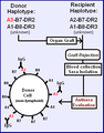
 Esquema diferenciación entre EICH y EICT.png 712 × 448;16 KB
-
 Esquema IPMA 3.jpg 853 × 496;62 KB
-
 Familias de PRRs.png 1,852 × 883;497 KB
-
 Fcvm-04-00048-g001.jpg 561 × 334;131 KB
-
 Fcvm-04-00048-g002.jpg 758 × 498;240 KB
-
 Fcvm-04-00048-g003-ko.jpg 965 × 732;230 KB
-
 Fcvm-04-00048-g003.jpg 965 × 732;456 KB
-
 Fcvm-05-00012-g001-ko.jpg 850 × 821;93 KB
-
 Fcvm-05-00012-g001.jpg 850 × 821;20 KB
-
 Fcvm-05-00012-g002.jpg 408 × 421;77 KB
-
 FcεR1.jpg 3,120 × 4,160;3.64 MB
-
 FebbreInfett.png 600 × 267;154 KB
-
 Fimmu-09-00585-g001-ko.jpg 965 × 502;276 KB
-
 Fimmu-09-00585-g001.jpg 965 × 502;293 KB
-
 Fimmu-09-00585-g002.jpg 968 × 1,117;492 KB
-
 Fimmu-09-01915-g0001.jpg 630 × 1,208;99 KB
-
 Fimmu-09-01915-g0002.jpg 630 × 1,094;110 KB
-
 Fimmu-09-01915-g003-ko.jpg 964 × 695;370 KB
-
 Fimmu-09-01915-g003.jpg 964 × 695;368 KB
-
 Fimmu-09-01915-g004.jpg 964 × 726;336 KB
-
 Fimmu-09-02948-g001.jpg 1,052 × 788;423 KB
-
 Fimmu-09-02948-g002.jpg 1,084 × 527;293 KB
-
 Fimmu-09-02948-g003.jpg 1,084 × 812;351 KB
-
 Fimmu-10-01699-g001.jpg 510 × 477;100 KB
-
 Fimmu-10-01699-g002.jpg 1,084 × 808;427 KB
-
 Fimmu-10-01699-g003.jpg 957 × 731;196 KB
-
 Fimmu-10-01699-g004.jpg 1,084 × 817;561 KB
-
 Fimmu-10-01699-g005.jpg 765 × 575;288 KB
-
 Fimmu-11-579250-g002.jpg 893 × 505;232 KB
-
 Fimmu-11-579250-g003.jpg 957 × 1,044;476 KB
-
 Fimmu-11-579250-g004-german version.png 893 × 686;368 KB
-
 Fimmu-11-579250-g004.jpg 893 × 686;319 KB
-
 First page of instructions for the inoculation of patients. Wellcome M0010782.jpg 2,771 × 4,027;1.51 MB
-
 Flow Cytometery IA.png 559 × 574;98 KB
-
 Fluorescence Assisted Cell Sorting (FACS) A.jpg 2,028 × 1,593;629 KB
-
 Fluorescence Assisted Cell Sorting (FACS) B.jpg 2,028 × 1,593;626 KB
-
 Fluorescently labeled antibodies.png 491 × 965;78 KB
-
 Fluorescenčně značené protilátky.jpg 491 × 965;170 KB
-
 Formació del precipitat del complex Ag-Ac.png 575 × 592;85 KB
-
 From paper4 2.png 730 × 510;23 KB
-
 From sec4 1.png 826 × 572;28 KB
-
 Fuentes de precursores hematopoyéticos.jpg 4,320 × 3,240;829 KB
-
 Genetic regulatory mechanisms affected by immunosenescence.jpg 800 × 311;166 KB
-
 Gliadin-immuno-innate.PNG 648 × 174;7 KB
-
 Gràfic Radi circumeferència- Ag.png 371 × 231;15 KB
-
 Gs4 sugar all.png 804 × 922;193 KB
-
 HAV IgM and IgG Test Results.jpg 1,920 × 1,080;511 KB
-
 HBsAg ELISA final reaction ready for reading.jpg 4,160 × 2,340;1.03 MB
-
 HerrmannMartin.jpg 800 × 1,069;1.04 MB
-
 HEV IgM and IgG Test Device.jpg 2,340 × 4,160;2.38 MB
-
 Hipotesistrasposon-gl.jpg 969 × 1,280;142 KB
-
 Historia de los transplantes.jpg 541 × 746;158 KB
-
 HIV ELISA final reaction in Microtiter plate wells for reading.jpg 2,340 × 4,160;2.74 MB
-
 Homeostasis del eritrocito y la hemoglobina.png 1,361 × 1,814;918 KB
-
 Hook effect.png 1,800 × 1,200;142 KB
-
 Human apoptosome ribbon rendering.png 640 × 516;301 KB
-
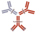 Humanisation.png 338 × 300;38 KB
-
Hy-Ձեռքբերովի իմունիտետ (Adaptive immune system).ogg 3分9秒;7.65 MB
-
 IBALT of mice.png 1,768 × 1,046;1.83 MB
-
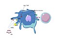
 IgE-und-Mastzellen.png 645 × 1,300;276 KB
-
 IgG.Opsonization.png 1,486 × 936;1.11 MB
-
 IgG1 vs IgG4 configuration.png 376 × 242;24 KB
-
 IgG4-Autoimmun-Organe.svg 2,314 × 1,609;2.23 MB
-
 IL1a Crystal Structure.png 851 × 730;126 KB
-
-
 ImdPathway Sept2019.jpg 448 × 510;60 KB
-
 Immkomp.jpg 432 × 446;137 KB
-
 Immuhistokemi.png 553 × 290;7 KB
-
 Immun resp.jpg 1,748 × 1,240;454 KB
-
 Immun-Organe-tr.png 434 × 586;83 KB
-
 Immun-Organe.png 434 × 586;91 KB
-

-
 Immune Figure 6.jpg 815 × 401;81 KB
-
 Immune modules 2.pdf 760 × 741;283 KB
-
 Immune modules.jpg 444 × 444;102 KB
-
 Immune repression by RIOK1.jpg 641 × 289;62 KB
-
 Immune response.jpg 1,136 × 704;75 KB
-
 Immune responses elicited by SARS-CoV-2 mRNA vaccines.webp 3,910 × 2,684;1.27 MB
-
 Immune22.gif 304 × 249;28 KB
-
 Immunhistokemi.png 314 × 236;4 KB
-
 Immunitat (medicina).png 1,412 × 525;51 KB
-
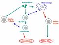
 Immunite lente et rapide.png 432 × 387;12 KB
-
 Immunity (medicine).срп.png 1,412 × 525;35 KB
-
 Immunity-ja.png 2,823 × 1,049;160 KB
-
 Immunity.png 1,412 × 525;20 KB
-
 Immunity.svg 1,324 × 492;15 KB
-
 Immuno 020.JPG 4,000 × 3,000;3.6 MB
-
 Immunocomplexes.png 1,016 × 800;79 KB
-
 Immunoinformatics.png 1,381 × 802;20 KB
-
 Immunologist Michel Sadelain.jpg 6,192 × 4,128;9.04 MB
-
 Immunology in the heart of Science.png 1,024 × 1,024;638 KB
-
 Immunomedia logo.png 6,096 × 2,032;1.18 MB
-
 Immunosenescence.jpg 3,543 × 2,362;1.46 MB
-
 Immunosoppress.png 400 × 277;102 KB
-
 Immunostaining Image.jpg 960 × 960;184 KB
-
 Immunostimol.png 400 × 276;104 KB
-
 ImmunSist.png 800 × 610;486 KB
-
 Imunita.jpg 1,412 × 525;53 KB
-
 Incandescence.png 2,048 × 2,048;2.95 MB
-
 Incomplete vs. Complete Clonal Deletion.png 1,322 × 726;197 KB
-
 Inflammaging.jpg 4,320 × 3,240;969 KB
-
 Inmunosenescencia.jpg 4,320 × 3,240;1,008 KB
-
 Instruments used for vaccination in the mid 19th century. Wellcome M0014876.jpg 3,709 × 2,833;1.7 MB
-
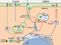
 Intestine immunology scheme.jpg 1,435 × 1,448;237 KB
-
 IRF8 in host response.png 720 × 915;96 KB
-
 IRGs.jpg 756 × 958;112 KB
-
 ITSU team structure.jpg 700 × 408;33 KB
-
 J558L (Mouse B Myeloma) Cell Line.jpg 335 × 248;98 KB
-
 JAK.STAT pathway.png 958 × 669;55 KB
-
 Killingt.JPG 457 × 356;19 KB
-
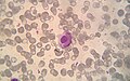 LECell.jpg 1,274 × 798;817 KB


.png)


.png)


;_5_-_granule;_6_-_mastocit;_7_-_novoformirani_medijatori_(prostaglandini,_leukotrieni).png)


_A.jpg)
_B.jpg)


_Cell_Line.jpg)


.png)
.%D1%81%D1%80%D0%BF.png)