Category:Microglia
Naar navigatie springen
Naar zoeken springen
glial cell located throughout the brain and spinal cord  Travmatik beyin hasarından önce sıçan korteksinden istirahat halindeki mikroglia (HRP ile lektin boyaması) | |||||
| Media uploaden | |||||
| Is een | |||||
|---|---|---|---|---|---|
| Subklasse van | |||||
| Onderdeel van | |||||
| |||||
Media in categorie "Microglia"
Deze categorie bevat de volgende 92 bestanden, van in totaal 92.
-
-
-
-
-
-
-
-
-
-
-
-
-
-
-
-
-
-
-
 Blausen 0870 TypesofNeuroglia ukr.png 2.323 × 1.822; 1,36 MB
-
 Blausen 0870 TypesofNeuroglia-sv.png 2.323 × 1.822; 1,77 MB
-
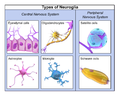 Blausen 0870 TypesofNeuroglia.png 2.500 × 2.000; 1,7 MB
-
-
-
-
Cellular-Basis-of-Pineal-Gland-Development-Emerging-Role-of-Microglia-as-Phenotype-Regulator-pone.0167063.s004.ogv 41 s, 1.920 × 1.080; 2,76 MB
-
Cellular-Basis-of-Pineal-Gland-Development-Emerging-Role-of-Microglia-as-Phenotype-Regulator-pone.0167063.s005.ogv 1 min 5 s, 1.280 × 720; 4,06 MB
-
Cellular-Basis-of-Pineal-Gland-Development-Emerging-Role-of-Microglia-as-Phenotype-Regulator-pone.0167063.s006.ogv 32 s, 1.920 × 1.080; 4,55 MB
-
Critical-Endothelial-Regulation-by-LRP5-during-Retinal-Vascular-Development-pone.0152833.s001.ogv 3,0 s, 512 × 512; 765 kB
-
Critical-Endothelial-Regulation-by-LRP5-during-Retinal-Vascular-Development-pone.0152833.s002.ogv 19 s, 512 × 512; 1,53 MB
-
Critical-Endothelial-Regulation-by-LRP5-during-Retinal-Vascular-Development-pone.0152833.s003.ogv 5,9 s, 512 × 512; 1,64 MB
-
-
Critical-Endothelial-Regulation-by-LRP5-during-Retinal-Vascular-Development-pone.0152833.s005.ogv 9,4 s, 512 × 512; 820 kB
-
Critical-Endothelial-Regulation-by-LRP5-during-Retinal-Vascular-Development-pone.0152833.s006.ogv 6,3 s, 512 × 512; 1.001 kB
-
Enhancement-of-Chemotactic-Cell-Aggregation-by-Haptotactic-Cell-To-Cell-Interaction-pone.0154717.s004.ogv 4,8 s, 754 × 750; 12,41 MB
-
-
Enhancement-of-Chemotactic-Cell-Aggregation-by-Haptotactic-Cell-To-Cell-Interaction-pone.0154717.s006.ogv 4,8 s, 754 × 750; 10,43 MB
-
-
-
-
In-Situ-Dividing-and-Phagocytosing-Retinal-Microglia-Express-Nestin-Vimentin-and-NG2-In-Vivo-pone.0022408.s001.ogv 5,0 s, 1.245 × 1.245; 396 kB
-
In-Situ-Dividing-and-Phagocytosing-Retinal-Microglia-Express-Nestin-Vimentin-and-NG2-In-Vivo-pone.0022408.s002.ogv 9,0 s, 1.245 × 1.245; 1,14 MB
-
-
-
-
-
-
-
-
-
-
-
-
-
-
-
-
-
 Microglia and neurons.jpg 1.600 × 1.200; 258 kB
-
 Microglia Elisa Iba1 cultiu 3 placa 3B cd8.tif 1.376 × 2.066; 16,23 MB
-
 Microglia in ischemic stroke.png 1.189 × 950; 1,67 MB
-
 Microglia lamelipodio.png 550 × 1.176; 426 kB
-
 Microglia phagoptosis of neuron cell.png 900 × 600; 22 kB
-
 Microglia Tactismo.PNG 1.126 × 1.872; 271 kB
-
 Microglia.svg 744 × 1.052; 19 kB
-
Microglial-Interactions-with-Synapses-Are-Modulated-by-Visual-Experience-pbio.1000527.s016.ogv 2,4 s, 516 × 417; 288 kB
-
Microglial-Interactions-with-Synapses-Are-Modulated-by-Visual-Experience-pbio.1000527.s017.ogv 2,6 s, 630 × 612; 749 kB
-
Microglial-Interactions-with-Synapses-Are-Modulated-by-Visual-Experience-pbio.1000527.s018.ogv 2,6 s, 797 × 729; 899 kB
-
Microglial-Interactions-with-Synapses-Are-Modulated-by-Visual-Experience-pbio.1000527.s019.ogv 1,2 s, 625 × 575; 349 kB
-
Microglial-Interactions-with-Synapses-Are-Modulated-by-Visual-Experience-pbio.1000527.s020.ogv 1,2 s, 918 × 840; 514 kB
-
Microglial-Interactions-with-Synapses-Are-Modulated-by-Visual-Experience-pbio.1000527.s021.ogv 1,2 s, 974 × 934; 866 kB
-
-
-
-
-
-
-
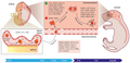 Microgliogenesis at a glance.png 4.338 × 2.089; 2,68 MB
-
 Mikroglej 1.jpg 480 × 357; 20 kB
-
-
-
-
-
-
 Purinergic signalling Microglia.jpg 843 × 621; 247 kB
-
Selective-targeting-of-microglia-by-quantum-dots-1742-2094-9-22-S1.ogv 5,1 s, 288 × 227; 115 kB
-
 Step-by-step guide for analyzing microglia phenotypes.png 1.978 × 2.656; 7,89 MB
-
-
-
-
TGF-signaling-in-the-brain-increases-with-aging-and-signals-to-astrocytes-and-innate-immune-cells-1742-2094-7-62-S5.ogv 5,1 s, 1.028 × 1.024; 5,49 MB
-
 WGA labelled microglial cells.tif 1.376 × 1.032; 4,06 MB
-
Zigzag-Turning-Preference-of-Freely-Crawling-Cells-pone.0020255.s009.ogv 15 s, 960 × 480; 320 kB


