Category:Molecular biology
Przejdź do nawigacji
Przejdź do wyszukiwania
branch of biology that deals with the molecular basis of biological activity  | |||||
| Prześlij plik multimedialny | |||||
| Jest to | |||||
|---|---|---|---|---|---|
| Podklasa dla | |||||
| Część | |||||
| Składa się z |
| ||||
| |||||

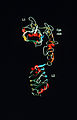
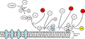
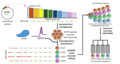
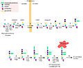
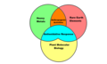
English: Molecular biology is the study of biology at a molecular level. The field overlaps with other areas of biology, particularly genetics and biochemistry. Molecular biology chiefly concerns itself with understanding the interactions between the various systems of a cell, including the interrelationship of DNA, RNA and protein synthesis and learning how these interactions are regulated.
Podkategorie
Poniżej wyświetlono 76 spośród wszystkich 76 podkategorii tej kategorii.
*
B
- Bacterial one-hybrid system (13 plików)
C
- Chromatin immunoprecipitation (21 plików)
- Cre-Lox recombination (9 plików)
D
E
F
G
- Genetic circuit (6 plików)
- GUS reporter system (5 plików)
H
I
M
- Macromolecular complex analysis (41 plików)
- Media from BMC Molecular Biology (20 plików)
- Media from EMBO Reports (5 plików)
- Media from Frontiers in Molecular Neuroscience (37 plików)
- Media from Molecular Brain (52 pliki)
- Media from Molecular Cancer (48 plików)
- Media from Molecular Pain (8 plików)
- Media from Molecular Vision (2 pliki)
- Media from The EMBO Journal (7 plików)
- Multiple displacement amplification (4 pliki)
N
O
- Open reading frames (20 plików)
- Optical tweezers (90 plików)
P
S
- Screening and selection (8 plików)
- Site-directed mutagenesis (52 pliki)
- Southern blot (13 plików)
T
- Two-hybrid system techniques (29 plików)
W
- WikiPathways (39 plików)
Strony w kategorii „Molecular biology”
W tej kategorii jest tylko jedna strona.
Pliki w kategorii „Molecular biology”
Poniżej wyświetlono 200 spośród wszystkich 299 plików w tej kategorii.
(poprzednia strona) (następna strona)-
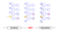
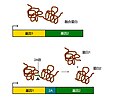 (zh)2A peptide Working Mechanism.jpg 593 × 493; 125 KB
-
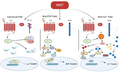
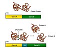 2A peptide Working Mechanism.jpg 593 × 493; 129 KB
-
 41598 2018 20107 Fig3 HTML.jpg 660 × 315; 167 KB
-
 4nqo1.gif 521 × 119; 6 KB
-
 4nqo2.png 784 × 194; 60 KB
-
 Acquaporina subunità porocanale.png 986 × 492; 27 KB
-
 Affinity Chromatography.jpg 580 × 800; 161 KB
-
 Affymetrix GeneChip.jpg 750 × 600; 312 KB
-
 Ahelix-FMO-Facio.jpg 1073 × 821; 105 KB
-
 Annotated structure of eRF1.jpg 600 × 361; 81 KB
-
 Apotosis.jpg 1302 × 520; 157 KB
-
 Application of MNAse.png 959 × 540; 194 KB
-
 Arnmensajero1.png 283 × 212; 28 KB
-
 ASO dot blot inverso.png 1310 × 786; 42 KB
-
 ASO dot blot.png 1304 × 864; 38 KB
-
 Balizas moleculares.png 960 × 720; 11 KB
-
 Bilal Djeghout Laboratory.jpg 1966 × 2048; 361 KB
-
 Binding.png 888 × 572; 22 KB
-
 Biological information flow.tif 1150 × 461; 2,57 MB
-
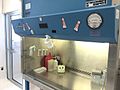 Biological Safety Cabinet (Class II, Type A2) Front view.jpg 1469 × 1102; 420 KB
-
 Biological Safety Cabinet (Class II, Type A2) Side view.jpg 1469 × 1102; 503 KB
-
 Biological Safety Cabinet (Class II, Type A2) Telstar front view.jpg 2048 × 1536; 731 KB
-
 Canonical and non-canonical Wnt signaling pathways.png 3620 × 2214; 1,06 MB
-
 Cascada JNK.jpg 2001 × 1501; 194 KB
-
 Cassettes.png 960 × 720; 42 KB
-
 CDNA-library-amplification.png 2955 × 1419; 159 KB
-
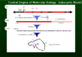 Central Dogma Model.png 800 × 557; 53 KB
-
 Choosing a cloning method.png 2625 × 1425; 144 KB
-
 Chromatogramme.jpg 583 × 768; 51 KB
-
 ColonneADN.JPG 153 × 359; 4 KB
-
 ColonneElution.JPG 153 × 359; 6 KB
-
 ConceptLarge.jpg 97 × 380; 28 KB
-
 Condensation3.png 2852 × 791; 279 KB
-
 Consumo excesivo de alimentos altos en Triacilgliceroles.jpg 897 × 701; 158 KB
-
 COREcassette.PNG 960 × 720; 43 KB
-
 Courtney 2008.jpg 786 × 685; 52 KB
-
 CreLoxP.jpg 320 × 316; 13 KB
-
 Crystal-by-example-illustrating-how-a-helix-might-be-formed.jpg 600 × 514; 82 KB
-
 CSIRO ScienceImage 2082 Cell Culture.jpg 2657 × 1976; 5,15 MB
-
 CSIRO ScienceImage 2738 First Three Domains of Insulin.jpg 1712 × 2657; 4,25 MB
-
 Ctd role .png 278 × 478; 22 KB
-
 Deconvoluted ESMS.jpg 1648 × 1123; 71 KB
-
 Dien bien nhanh.JPG 478 × 709; 64 KB
-
 Dieu hoa di hinh lap the-01.png 9542 × 6723; 652 KB
-
 Digestión, absorción y metabolismo de ácidos grasos.jpg 1018 × 814; 215 KB
-
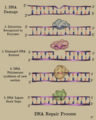 DNA repair.png 1080 × 1350; 627 KB
-
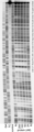
 DNA-Leiter.jpg 404 × 802; 55 KB
-
 DNaseI footprint.png 162 × 626; 79 KB
-
 DogmaCentrale.svg 728 × 239; 33 KB
-
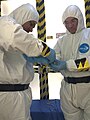 Donning.jpg 1102 × 1469; 440 KB
-
 Droga alternatywna.png 759 × 350; 66 KB
-
 Droga klasyczna.png 725 × 405; 74 KB
-
 Ecoli human compare.jpg 675 × 450; 51 KB
-
 EcoRV structure.png 1470 × 1757; 1,98 MB
-

-
 Electronic access control (BSL3 Lab) using magnetic swipe card.jpg 2048 × 1536; 702 KB
-

-
 Electronic access control (BSL3 Lab).jpg 2048 × 1536; 725 KB
-
 Epissage.png 565 × 222; 10 KB
-
 Epitelo-mezenchymální tranzice.gif 822 × 214; 36 KB
-
 Erkennungssequenzen von Restriktionsenzymen.jpg 764 × 331; 22 KB
-
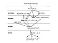
 EsquemaBiologiaMolecular.png 576 × 354; 599 KB
-
 Estructura ORC.png 399 × 216; 21 KB
-
 Experimental technique Dip-c.jpg 1771 × 338; 101 KB
-
 Experimento-pulso-caza.jpg 500 × 272; 39 KB
-
 Extraction Chamber of Molecular Laboratory.jpg 4000 × 2250; 2,39 MB
-
 Extrapolation based Molecular Systems biology GRana 09.jpg 960 × 720; 73 KB
-
 Extrapolation based Molecular Systems biology Rana 09.jpg 960 × 720; 73 KB
-
 Extrapolation Based Molecular Systems Biology Rana CWRU.tiff 960 × 720; 589 KB
-
 Faire scheme.png 3000 × 2300; 1,78 MB
-
 Fedg.png 1086 × 817; 363 KB
-
 Fig2.Recombination patterns.png 960 × 720; 12 KB
-
 Figura Wiki.png 720 × 504; 206 KB
-
 Figure 1 NAPPA.png 1363 × 470; 69 KB
-
 Figure 2 PISA.png 1315 × 374; 61 KB
-
 Figure 3 puromycin2.png 1415 × 469; 93 KB
-
 Figure 4 nano well.png 1390 × 403; 62 KB
-
 Figure 5 DAPA.png 1392 × 628; 60 KB
-
 Figure final 3.jpg 3071 × 2457; 491 KB
-
 Finite Element Model.jpg 793 × 634; 41 KB
-
 Finite model.jpg 1050 × 734; 53 KB
-
 Fish Egg Diagram (1).jpg 960 × 720; 38 KB
-
 Fish Egg.jpg 960 × 720; 37 KB
-
 FlAsh Protein Modification.png 2161 × 676; 110 KB
-
 Functional Cloning.png 5500 × 800; 611 KB
-
 Genequant.jpg 1536 × 2048; 226 KB
-
 GESTALT workflow.png 1436 × 769; 107 KB
-
 GolgiTethersc.jpg 390 × 233; 28 KB
-
 Gpi synthesis.jpg 4187 × 3455; 1,79 MB
-
 Grafica de la PCR.jpg 528 × 600; 77 KB
-
 Heavy Metals vs REE vs Plant Molecular Biology.png 876 × 526; 51 KB
-
 HIVE Annotation Mapper Computation.png 927 × 885; 130 KB
-
 HIVE Heptagon Computation.png 1034 × 1161; 252 KB
-
 HIVE Hexagon Computation.png 1015 × 850; 185 KB
-
 HIVE Hexahedron Computation.png 969 × 1053; 221 KB
-
 HIVE IDBA-UD Computation.png 1102 × 885; 151 KB
-
 HIVE MAFFT Computation.png 1075 × 728; 174 KB
-
 HIVE Velvet Computation.png 1102 × 696; 121 KB
-
 Hmm necleotides 2.pdf 856 × 881; 49 KB
-
 Hmm nucleotide seq.pdf 1354 × 137; 18 KB
-
 Human genome to genes zh.png 1454 × 866; 386 KB
-
 Hydrophobic Mismatch.JPG 936 × 995; 164 KB
-
 Hypothesis Scarano-etal.1967.png 567 × 433; 19 KB
-
 Hêlicaza Helicase.png 150 × 448; 19 KB
-
 Hêlicaza tác động Helicase in action.png 200 × 448; 31 KB
-
 Initial model of chlororespiration.jpg 797 × 556; 29 KB
-
 Isoforms and sequence.png 676 × 620; 44 KB
-
 Jeewanu globules - Electron Imaging.png 342 × 446; 122 KB
-
 Journal.pone.0001604.g001 small.jpg 597 × 144; 26 KB
-

-
 Klonierung2.png 624 × 239; 16 KB
-
 La technologie BRET.png 1022 × 1162; 49 KB
-

-
 Latest understanding of chlororespiration (2002).jpg 937 × 527; 31 KB
-
 Lentiviral vector.png 961 × 467; 126 KB
-
 LH2 side.jpg 1048 × 904; 151 KB
-
 Matrix Metalloproteinases.png 728 × 434; 45 KB
-
 Mattress model.JPG 921 × 729; 92 KB
-
 MCR1erythro4TC.jpg 564 × 304; 41 KB
-
 Mechanism of Nonsense Mediated Decay.jpg 631 × 1155; 150 KB
-
 Mediator4TC.jpg 357 × 289; 41 KB
-
 MediatorPolII4TC.jpg 326 × 224; 34 KB
-
 MediatorPolTF4TC.jpg 603 × 270; 69 KB
-
 MediatorSpline4TC.jpg 194 × 240; 21 KB
-
 MediatorStrucMod4TC.jpg 271 × 168; 21 KB
-
 Membrane lipids.png 579 × 607; 10 KB
-
 Membrane potential development.jpg 1619 × 518; 79 KB
-
 Membrane potential ions (id).jpg 550 × 400; 186 KB
-
 MirrorImagePhageDisplay.png 1775 × 691; 150 KB
-
 MNAse based sequencing.png 360 × 622; 94 KB
-
 MNase based sequencing.png 360 × 622; 94 KB
-
 Mnase image.png 360 × 622; 84 KB
-
 Molecular response after nerve injury.pdf 962 × 806; 243 KB
-
 Molecular response after nerve injury.png 1926 × 1613; 550 KB
-
 Molekulaarbioloogia põhidogma.svg 794 × 300; 7 KB
-

-

-

-
 Musclon2.jpg 556 × 442; 29 KB
-
 MutHPvuII.png 1075 × 498; 467 KB
-
 Mô hình enzyme.svg 512 × 512; 6 KB
-
 Na-K pump cycle.jpg 800 × 457; 108 KB
-
 Na-K pump cycle.png 800 × 457; 156 KB
-
 NASBA fase 1.jpg 1508 × 929; 69 KB
-
 NASBA fase 2.jpg 1495 × 1135; 115 KB
-
 Nearest-Neighbor-seq-freq XpY.png 1665 × 886; 121 KB
-
 Neocentromere Formation .jpg 2917 × 3508; 1,71 MB
-
 Neubauer improved with cells.jpg 1280 × 960; 669 KB
-
 Neural progenitor cells.png 3893 × 939; 637 KB
-
 Neuraminidase2.jpg 785 × 662; 349 KB
-
 Nick translation.svg 592 × 1029; 29 KB
-
 NLRP3.png 1460 × 976; 645 KB
-
 NMD - Nonsense-mediated decay.png 7292 × 4965; 709 KB
-

-
 Nrm2503-f2.jpg 655 × 440; 118 KB
-
 Nuclear integrity and genome stability in normal and HGPS cells.jpg 600 × 723; 64 KB
-
 Outline of no-SCAR recombineering methods final 2.png 2400 × 2810; 25,74 MB
-
 PAPRs in use 01.jpg 1280 × 853; 100 KB
-
 Parik1.jpg 680 × 552; 65 KB
-
 Parik2.1.jpeg 388 × 636; 29 KB
-
 Parik3.jpeg 623 × 642; 70 KB
-
 Parik4.jpg 706 × 334; 55 KB
-
 Pathways of glucolysis.png 1366 × 768; 90 KB
-
 PCR es.png 300 × 675; 26 KB
-
 Penetrance.pdf 1239 × 1752; 51 KB
-
 Penetrance1.0.pdf 1752 × 1239; 49 KB
-
 Penetrance2.pdf 1239 × 1752; 56 KB
-
 PenetranceVE.pdf 1752 × 1239; 56 KB
-
 Perfil de exitación y emision de DAPI.jpg 252 × 220; 13 KB
-
 Peroxisome Dynamics-Molecular Players,Mechanisms, and (Dys)functions.pdf 1250 × 1650, 25 stron; 375 KB
-
 PETworkflow.png 1061 × 797; 75 KB
-
 PGEX-3X cloning vector.png 356 × 354; 10 KB
-
 Philadelphia chromosome detection.jpg 563 × 669; 133 KB
-
 Physiology.. .png 427 × 387; 25 KB
-
 Pipette tip over tube.jpg 2361 × 3305; 623 KB
-
 Piskacek TF1.jpg 8080 × 3456; 1,57 MB
-
 PL Mykowirusy – wirusy infekujące grzyby J. Kamiński.pdf 1239 × 1754, 56 stron; 1,82 MB
-
 PL Wstępna charakterystyka bakteriofaga Serratia φOS10 J. Kamiński.pdf 1239 × 1752, 101 stron; 2,13 MB
-
 Plant cell structure svg vacuole (id).jpg 649 × 475; 156 KB
-
 Plant-cell-sucrose-gradient-fractions.jpg 2988 × 5312; 1,74 MB
-
 Polysomesleft.jpg 402 × 518; 45 KB
-
 PomBase infographic.jpg 1591 × 2250; 608 KB
-
 Post063a - Flickr - NOAA Photo Library.jpg 1800 × 1200; 2,1 MB
-
 Powerlaw HI II 14.png 536 × 391; 24 KB
-
 Preparing PCR reaction.jpg 4128 × 2322; 1,17 MB
-
 Principle of competent cell preparation 1.png 2149 × 1627; 421 KB
-
 Principle of competent cell preparation 2.png 2154 × 1627; 436 KB
-
 Proces de la PCR.jpg 300 × 675; 81 KB
-
 PromotorsK01589 logo 1.pdf 1050 × 1050; 51 KB
-
 Protein co-localization on microtubules.png 2160 × 2160; 8,09 MB
-
 Protein con duong nhanh.JPG 600 × 364; 175 KB
-
 Proton trap.jpg 690 × 409; 60 KB
-
 Proves de la TaqMan.jpg 650 × 167; 23 KB
-
 ProximityAssay14TC.jpg 269 × 92; 8 KB
-
 ProximityAssay24TC.jpg 270 × 231; 15 KB
-
 ProximityAssay34TC.jpg 269 × 229; 13 KB
-
 ProximityAssay44TC.jpg 269 × 238; 21 KB
-
 ProximityAssay5Cell4TC.jpg 211 × 216; 11 KB
-
 PROYECTO EDUCATIVO wikilibros.jpg 960 × 540; 76 KB
-
 Pymol nahled.png 859 × 765; 183 KB
2A_peptide_Working_Mechanism.jpg)


_Front_view.jpg)
_Side_view.jpg)
_Telstar_front_view.jpg)


_using_magnetic_swipe_card.jpg)
_using_personal_identification_number_(PIN).jpg)
.jpg)


.jpg)


.jpg)


.jpg)


.jpg)


