Category:Nervous system
Prijeđi na navigaciju
Prijeđi na pretraživanje
part of an animal that coordinates actions and senses  | |||||
| Postavi datoteku | |||||
| Zvučni zapis izgovora | |||||
|---|---|---|---|---|---|
| Jest |
| ||||
| Dio klase |
| ||||
| Dio |
| ||||
| Materijal | |||||
| Sastoji se od | |||||
| |||||
Potkategorije
Ova kategorija ima sljedećih 27 potkategorija, od ukupno 27.
A
B
- Biologische Untersuchungen (21 F)
C
- Nervous system components (15 F)
L
- Leptin (obesity protein) (22 F)
M
N
- Neural cell adhesion molecules (13 F)
- Neural inhibition (10 F)
O
P
S
- Superior olivary complex (2 F)
T
Mediji u kategoriji »Nervous system«
Prikazano je 198 datoteka u ovoj kategoriji, od njih ukupno 198.
-
 Aferencias para SNC.png 1.485 × 347; 38 KB
-
 Aferentní a eferentní nervová dráha.jpg 1.941 × 1.113; 172 KB
-
 Afferent and efferent neurons zh.png 461 × 283; 50 KB
-
 Allgemeine Anatomie und Physiologie des Nervensystems (1903) (18104074332).jpg 1.555 × 3.154; 767 KB
-
 Allgemeine Anatomie und Physiologie des Nervensystems (1903) (18108488551).jpg 2.736 × 1.598; 585 KB
-
 Alteration theory of nerve impulse propagation Wellcome L0002237EA.jpg 2.564 × 730; 103 KB
-
 Alteration theory of nerve impulse propagation Wellcome L0002237EB.jpg 1.782 × 1.064; 273 KB
-

-
-
 Anatomy; pons, 1844 Wellcome M0014312.jpg 3.288 × 3.296; 1,5 MB
-
Animação ampola 2.webm 13 s, 820 × 720; 1,97 MB
-
Animação ampola.webm 9,2 s, 820 × 720; 199 KB
-
 Apparatus for electrical stimulation of nerves Wellcome M0011390.jpg 4.081 × 2.588; 1,94 MB
-
 Apparatus used in experiments in curare, C. Bernard Wellcome M0014744.jpg 2.534 × 4.300; 4,49 MB
-
 Arteries and veins of the brain. Wellcome L0000989.jpg 1.124 × 1.646; 789 KB
-
 Ativação e inibição na célula ciliada.png 1.024 × 925; 60 KB
-
 B. Lewis, Cortical cells in sheep. Wellcome L0001988.jpg 1.745 × 1.087; 807 KB
-
 Bastonetes células bipolares e celulas ganglionares centro-periferia.png 1.461 × 787; 375 KB
-
 Bastonetes e células bipolares no claro e no escuro com glutamato.png 1.133 × 937; 124 KB
-
 Bastonetes e células bipolares no claro e no escuro eletrofisiologia.png 1.360 × 768; 91 KB
-
 Bastonetes e células bipolares no claro e no escuro simples.png 1.188 × 1.083; 98 KB
-

-
-
 Bergê maylînî ku.png 691 × 422; 103 KB
-
 Betz cells of premotor cerebral cortex. Wellcome L0001958.jpg 1.391 × 1.347; 376 KB
-
 Bidloo Ontleding 1690 10.jpg 1.200 × 3.608; 584 KB
-
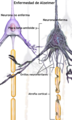 Blausen 0017 AlzheimersDisease-es.png 1.400 × 2.334; 2,47 MB
-
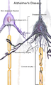 Blausen 0017 AlzheimersDisease.png 1.350 × 2.250; 8,69 MB
-
 Brain and spinal cord; six figures showing various portions. Wellcome V0008407.jpg 2.162 × 3.484; 3,69 MB
-
 Cajal (1888) firt drawing of nervous system.gif 150 × 92; 14 KB
-
 Canais de sodio e potassio despolarizacao hiperpolarizacao.png 1.418 × 770; 140 KB
-
 Celula ciliada cilio inclinado influxo efluxo potassio canais abertos.png 1.047 × 1.168; 112 KB
-

-
-
 Celula ciliada cilio inclinado influxo efluxo potassio influxo calcio.png 1.047 × 1.168; 131 KB
-
 Celula ciliada cilio inclinado influxo potassio canais abertos.png 1.047 × 1.168; 75 KB
-
 Celula ciliada cilio inclinado influxo potassio.png 1.047 × 1.168; 74 KB
-
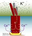
 Celula ciliada cilio inclinado perilinfa endolinfa.png 1.073 × 1.145; 75 KB
-
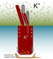
 Celula ciliada cilio inclinado.png 1.062 × 1.145; 60 KB
-
 Celula ciliada repouso.png 1.062 × 1.145; 55 KB
-
 Cerebral cortex of a child. Wellcome L0002012.jpg 1.004 × 1.904; 677 KB
-
 Cerebral cortex of adult cat. Wellcome L0002047.jpg 1.140 × 1.616; 573 KB
-
 Cilios inclinados receptores ancorados.png 1.812 × 649; 102 KB
-
-
 Complete neuron cell diagram tr.svg 819 × 596; 271 KB
-
 Cristalino e musculo ciliar contraido relaxado.png 655 × 783; 50 KB
-
 Cílios inclinados membrana esticada afrouxada.png 1.076 × 669; 65 KB
-
 Cóclea desespiralada 1.png 1.742 × 274; 35 KB
-
 Cóclea desespiralada 2.png 1.742 × 397; 47 KB
-
 Cóclea desespiralada 3.png 1.742 × 506; 54 KB
-
 Cóclea desespiralada 4.png 1.742 × 451; 59 KB
-
 Cóclea desespiralada 5.png 1.548 × 416; 198 KB
-
 De dermatomerie bij de hagedis (lacerta viridis) (1917) (20657060270).jpg 1.522 × 2.840; 1.015 KB
-
 De la structure du cerveau... Wellcome M0014439.jpg 2.952 × 3.680; 4,54 MB
-
 Demarexane ku.png 810 × 617; 206 KB
-
 Demaxê navberê ku.png 796 × 628; 251 KB
-
 Descartes' concept of the nerve. Wellcome L0001862.jpg 1.032 × 1.810; 571 KB
-
 Descartes; Coordination of muscle and visual mechanisms. Wellcome L0002392.jpg 1.172 × 1.630; 770 KB
-
 Descartes; Diagram of ventricles of brain. Wellcome L0008517.jpg 1.200 × 1.642; 586 KB
-
 Descartes; Quelle est la fabrique de ses nerfs. Wellcome M0014443.jpg 2.675 × 3.974; 967 KB
-
 Descartes; The Nervous System Wellcome L0002432.jpg 1.702 × 1.118; 984 KB
-
 Descartes; The Nervous System. Diagram of the brain Wellcome L0001371.jpg 1.746 × 1.096; 602 KB
-
 Descartes; The Nervous System. Diagram of the brain Wellcome L0006584.jpg 5.787 × 3.018; 2,84 MB
-
 Descartes; The path of burning pain. Wellcome M0014440.jpg 3.134 × 3.456; 3,86 MB
-
 Die Medusen (9492023901).jpg 2.232 × 3.088; 601 KB
-
 Die Medusen (9492025275).jpg 2.232 × 3.088; 748 KB
-
 Die Medusen (9492028437).jpg 2.232 × 3.088; 620 KB
-
 Die Medusen (9492034457).jpg 2.232 × 3.088; 821 KB
-
 Die Medusen (9492037097).jpg 2.232 × 3.088; 773 KB
-
 Die Medusen (9494825878).jpg 2.232 × 3.088; 792 KB
-
 Die Medusen (9494826800).jpg 2.232 × 3.088; 538 KB
-
 Die Medusen (9494829152).jpg 2.232 × 3.088; 537 KB
-
 Die Medusen (9494830360).jpg 2.232 × 3.088; 638 KB
-
 Die Medusen (9494831414).jpg 2.232 × 3.088; 581 KB
-
 Die Medusen (9494832666).jpg 2.232 × 3.088; 667 KB
-
 Die Medusen (9494835304).jpg 2.232 × 3.088; 728 KB
-
 Die Medusen (9494837596).jpg 2.232 × 3.088; 574 KB
-
 Distribution of spinal roots and muscles Wellcome L0001973.jpg 1.557 × 1.166; 401 KB
-
 Dor e temperatura.png 1.468 × 908; 144 KB
-
 Electrical state of muscle fibres Wellcome L0001987.jpg 1.990 × 952; 801 KB
-
 Electrical state of muscle Wellcome L0001986.jpg 2.066 × 920; 572 KB
-
 Encephalitis, in Specilegium anatomicum Wellcome L0005443.jpg 1.162 × 1.580; 827 KB
-
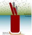
 Epitelio ciliado endolinfa perilinfa.png 1.425 × 849; 53 KB
-
 Esquema do Sistema Nervoso de Sipuncula.png 425 × 342; 16 KB
-
 Esquemes sistema nerviós.PNG 208 × 579; 59 KB
-
 Estríola.png 1.425 × 369; 49 KB
-
 Evolucion snc.PNG 1.450 × 520; 69 KB
-
 Experiments on animal electricity, 19th Century Wellcome L0001985.jpg 1.506 × 1.258; 624 KB
-
 Faisceau acoustique Reil.png 584 × 672; 635 KB
-
 Fibre net of Joseph von Gerlach Wellcome L0002013.jpg 907 × 2.047; 716 KB
-
 Figure expliquant la Théorie du Gate Control.pdf 1.239 × 1.754; 391 KB
-
 Fneur-10-00574-g001.jpg 765 × 543; 277 KB
-
 Función MT5-MMP.png 1.582 × 1.054; 324 KB
-
 Golgi's drawing of nervous system.gif 150 × 139; 22 KB
-
 Groeikegel.jpg 355 × 238; 15 KB
-
 Guainanodoranvier.jpg 414 × 131; 10 KB
-
 Hermosto2.jpg 571 × 258; 21 KB
-
 Human figure, venous and nervous system, Persian, 18th C Wellcome L0012607.jpg 1.182 × 1.445; 734 KB
-
 Illustration of nervous system and nerve fibres Wellcome L0001861.jpg 1.192 × 1.614; 779 KB
-
Indução de movimentos dos olhos.webm 20 s, 1.280 × 720; 674 KB
-
 Inércia nos órgãos otolíticos.png 1.764 × 1.055; 190 KB
-
 J-M Charcot, Lecons sur les maladies du syst Wellcome L0029848.jpg 1.208 × 1.752; 862 KB
-
 J.M. Charcot, Lectures on the diseases of th Wellcome L0029868.jpg 1.278 × 1.618; 708 KB
-
 J.M. Charcot, Lectures on the diseases of th Wellcome L0029869.jpg 1.424 × 1.454; 751 KB
-
Janela redonda.webm 10 s, 1.280 × 720; 268 KB
-
 Jardine Naturalist's library Entomology Plate 4.jpg 1.833 × 2.933; 671 KB
-
 Kinocilium.jpg 1.388 × 1.295; 174 KB
-
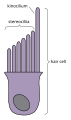 Kinocilium.svg 594 × 1.001; 9 KB
-
 Koendamademarê ku.png 848 × 590; 58 KB
-
 L'isthme de l'encéphale.png 566 × 595; 697 KB
-
 Le corps trapézoïde.png 891 × 576; 987 KB
-
 Limiar e intensidade do sinal sensorial.png 1.528 × 543; 49 KB
-
 Makrofagi 2.jpg 499 × 419; 24 KB
-
 Medula espinhal cervical com raízes.png 1.801 × 432; 72 KB
-
 Models for synaptogenesis.pdf 2.197 × 1.264; 26 KB
-
 Movimentos conjugados e disjuntivos eixo.png 1.062 × 726; 82 KB
-
 Movimentos conjugados e disjuntivos.png 1.385 × 701; 142 KB
-
 Narvisusteemi jaotus 2.png 494 × 326; 30 KB
-
 Narvisusteemijaotus.png 494 × 326; 30 KB
-
 Nerve Cells and Nerve Fibres Wellcome L0033035.jpg 3.685 × 4.810; 6,67 MB
-
 Nerve cells and nerve fibres. Wellcome L0002196.jpg 1.276 × 1.558; 740 KB
-
 Nerve cells surrounded by axone terminations. Wellcome L0001463.jpg 2.042 × 930; 340 KB
-
 Nerve cells. Wellcome L0001966.jpg 1.056 × 1.772; 691 KB
-
 Nervenzelle.jpg 3.507 × 2.550; 355 KB
-
 Nervenzelle.png 1.965 × 3.318; 533 KB
-
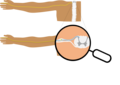 Nervo pelo braço.png 1.724 × 1.224; 95 KB
-
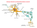
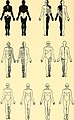
 Nervous system organization af.svg 601 × 482; 766 KB
-
 Nervous system organization en.svg 601 × 482; 767 KB
-
 Nervous system organization hu.svg 601 × 482; 759 KB
-
 Nervous system organization nl.jpg 950 × 760; 137 KB
-
 Nervous system organization-ar.svg 601 × 482; 929 KB
-
 Neurology, 19th century; cell of Schwann. Wellcome L0002015.jpg 1.212 × 1.577; 352 KB
-
 Neuromelanin in a neuron of the substantia nigra.jpg 855 × 552; 661 KB
-
 Nissl bodies in neurons of the spinal cord.jpg 1.200 × 900; 1,79 MB
-
Nistagmo.webm 12 s, 1.280 × 720; 2,24 MB
-
 NSdiagram af.svg 512 × 408; 9 KB
-
 NSdiagram unpathed.svg 744 × 1.052; 30 KB
-
 NSdiagram.png 622 × 513; 101 KB
-
 NSdiagram.svg 512 × 408; 4 KB
-
 NSDivisionsEstonian3.png 1.028 × 677; 77 KB
-
 Octopus nervous system.png 757 × 591; 60 KB
-
 Optic chiasm development.jpg 1.625 × 1.647; 1,03 MB
-
 Plancher du quatrième ventricule et tubercules quadrijumeaux.png 756 × 965; 1,41 MB
-
 Plano focal alvo visual distante.png 1.520 × 591; 52 KB
-
 Plano focal alvo visual proximo corrigido cristalino.png 1.989 × 608; 82 KB
-
 Plano focal alvo visual proximo.png 1.989 × 608; 52 KB
-
 Ponto focal na retina.png 1.954 × 591; 31 KB
-
 Potentiel action PA seuil.jpg 435 × 448; 24 KB
-
 Principles of Psychology (James) v1 p21.png 660 × 660; 145 KB
-
 Profundidade de foco aumentada.png 1.954 × 591; 40 KB
-
 Profundidade de foco curta.png 1.954 × 591; 50 KB
-
 Pyramidal tract in brain and cord Wellcome L0001977.jpg 1.022 × 1.818; 950 KB
-

-

-
-
 Refracao optica da visao diferentes angulos de chegada.png 2.000 × 572; 14 KB
-
 Refracao optica da visao.png 1.435 × 828; 42 KB
-
 Reizweiterleitung der Nervenzelle.jpg 2.480 × 3.508; 467 KB
-
 Retardo interaural.png 1.005 × 510; 47 KB
-
 Ro67-4853.png 1.559 × 763; 188 KB
-
 Ruffini ending.PNG 512 × 384; 5 KB
-
 Régulation de la pression artérielle.png 1.437 × 2.124; 1,39 MB
-
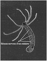 Réseau nerveux d'un cnidaire (hydre).jpg 2.604 × 3.363; 666 KB
-
Sacadas.webm 12 s, 1.280 × 720; 1,98 MB
-
 Sacral sympathetic.jpg 960 × 720; 113 KB
-
 Schema des Verlaufes..., 1892 Wellcome M0014444.jpg 2.233 × 4.806; 4,99 MB
-

-
 Schéma représentant dans son ensemble la voie motrice principale.png 649 × 1.060; 1,3 MB
-
 Schéma représentant, dans son ensemble, la voie sensitive centrale.png 706 × 1.081; 1,42 MB
-
Seguimento lento.webm 12 s, 1.280 × 720; 2,19 MB
-
 Semiologia de Sistema Nervioso.pdf 1.275 × 1.650, 9 stranice; 57 KB
-
 Sensory Pathways III.png 519 × 22; 7 KB
-
 Sites of punctures of 4th ventricle Wellcome M0011392.jpg 2.890 × 3.716; 2,08 MB
-
 Slide1BAB.JPG 960 × 720; 139 KB
-
 SNC esquematico.png 629 × 952; 34 KB
-
 Superior cervical ganglion 1.jpg 960 × 720; 101 KB
-
 Superior View of the Brain.jpg 521 × 457; 56 KB
-
 Synapse Illustration2 tweaked zh-CN.svg 862 × 555; 148 KB
-
 Tab 4, 'Observations on the ... nervous system' Wellcome L0050671.jpg 4.187 × 6.658; 3,85 MB
-
 Tab 5, 'Observations on the ... nervous system' Wellcome L0050673.jpg 4.238 × 6.802; 5,24 MB
-
 Tabs 37-39, 'Observations on the ... nervous system' Wellcome L0050674.jpg 4.323 × 6.778; 4,42 MB
-
 The brain as an organ of mind (1896) (14597249499).jpg 1.380 × 2.808; 756 KB
-
 The brain as an organ of mind (1896) (14597274759).jpg 1.408 × 1.148; 470 KB
-
 The brain as an organ of mind (1896) (14597279228).jpg 1.348 × 1.892; 409 KB
-
 The brain as an organ of mind (1896) (14597479887).jpg 1.684 × 2.512; 1,17 MB
-
 The brain as an organ of mind (1896) (14760929976).jpg 864 × 1.220; 214 KB
-
 The brain as an organ of mind (1896) (14783910615).jpg 1.400 × 2.068; 330 KB
-
 Timed views of a neve fibre. Wellcome L0002194.jpg 1.310 × 1.504; 1.014 KB
-

-
 Transducao.png 1.434 × 404; 21 KB
-
 Unmyelinated Nerve.jpg 615 × 345; 43 KB
-
 Vias ascendentes.png 1.352 × 752; 71 KB
-
-
-
-
-
 Wallerian degeneration of a nerve. Wellcome L0002019.jpg 1.228 × 1.520; 474 KB
-
 色質溶解Chromatolysis.jpg 500 × 388; 63 KB


_(18104074332).jpg)
_(18108488551).jpg)

_(17982113390).jpg)


_(19743465713).jpg)
_(20177724519).jpg)


_firt_drawing_of_nervous_system.gif)


_(20482916158).jpg)


_(1917)_(20657060270).jpg)


.jpg)
.jpg)
.jpg)
.jpg)
.jpg)
.jpg)
.jpg)
.jpg)
.jpg)
.jpg)
.jpg)
.jpg)
.jpg)


_v1_p21.png)

_(14574560428).jpg)
_(14758019571).jpg)


.jpg)


_(14597249499).jpg)
_(14597274759).jpg)
_(14597279228).jpg)
_(14597479887).jpg)
_(14760929976).jpg)
_(14783910615).jpg)

.jpg)


_(18169811105).jpg)
_(14574536559).jpg)