Category:Parasitology
Jump to navigation
Jump to search
branch of biology that studies parasites, their hosts, and the relationship between them .jpg) Паразитолог за работой | |||||
| മീഡിയ ചേർക്കുക | |||||
| ഉച്ചാരണ ശബ്ദം | |||||
|---|---|---|---|---|---|
| തരം |
| ||||
| ഇതിന്റെ ഉപവിഭാഗം | |||||
| ഇതിന്റെ ഭാഗം | |||||
| |||||
ഉപവർഗ്ഗങ്ങൾ
ഈ വർഗ്ഗത്തിൽ ആകെ 13 ഉപവർഗ്ഗങ്ങൾ ഉള്ളതിൽ 13 ഉപവർഗ്ഗങ്ങൾ, താഴെക്കൊടുത്തിരിക്കുന്നു.
*
B
D
H
I
J
- Journal of Parasitology (1 F)
P
- Parasitemia (2 F)
- Parasitic diseases (150 F)
Q
- Quantitative parasitology (9 F)
V
"Parasitology" എന്ന വർഗ്ഗത്തിലെ പ്രമാണങ്ങൾ
ഈ വർഗ്ഗത്തിൽ മൊത്തം 236 പ്രമാണങ്ങളുള്ളതിൽ 200 എണ്ണം താഴെ നൽകിയിരിക്കുന്നു.
(മുൻപത്തെ താൾ) (അടുത്ത താൾ)-
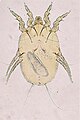
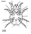 Acarus, female, ventral.png 3,308 × 3,615; 169 കെ.ബി.
-
 Adult Enterobius vermicularis.jpg 4,000 × 3,000; 5.18 എം.ബി.
-
 Aggregated distribution of parasites on hosts.svg 1,135 × 936; 17 കെ.ബി.
-
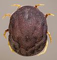
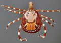 Amblyomma male dorsal.jpg 945 × 663; 481 കെ.ബി.
-
 Amblyomma-variegatum-female-male-dorsal.jpg 1,024 × 643; 291 കെ.ബി.
-
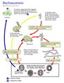
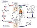
 Angiostrongyliasis lifecycle.png 618 × 694; 112 കെ.ബി.
-
 Angiostrongylus cantonensis life cycle 01.png 2,899 × 2,313; 570 കെ.ബി.
-
 Angiostrongylus costaricensis life cycle.png 2,927 × 2,298; 567 കെ.ബി.
-
 Anisakiasis life cycle.png 2,456 × 3,076; 686 കെ.ബി.
-
 Aparato reproductor de Duplaccessorius andinus visto en ampo oscuro.jpg 1,800 × 2,104; 1.76 എം.ബി.
-
 Archives de parasitologie (1900-1901) (19130575244).jpg 1,308 × 1,548; 1.04 എം.ബി.
-
 Argas-persicus-female-dorsal-ventral.jpg 1,116 × 692; 225 കെ.ബി.
-
 Baylisascaris procyonis life cycle CDC.tif 2,400 × 3,150; 21.65 എം.ബി.
-
 Baylisascaris procyonis life cycle.png 2,453 × 3,162; 774 കെ.ബി.
-
 Beschreibung eines neu-entdeckten Eingeweide-Wurms im menschlichen Körper (1802) (20367109545).jpg 2,944 × 3,070; 1.98 എം.ബി.
-
 Blastocystis, life cycle.jpg 742 × 530; 62 കെ.ബി.
-
 Book-title-Triatoma.png 4,757 × 1,885; 142 കെ.ബി.
-
 Bovicola louse ventral.jpg 1,176 × 2,072; 1.26 എം.ബി.
-
 Brugia malayi life cycle CDC.tif 3,150 × 2,400; 21.65 എം.ബി.
-
 Cephalopina-titillator-larva.jpg 1,024 × 561; 110 കെ.ബി.
-
 Cheyletiella-parasitivorax-mite.jpg 1,108 × 1,305; 471 കെ.ബി.
-
 Chorioptes-bovis-mite.jpg 1,316 × 1,609; 589 കെ.ബി.
-
 Chrysomya adult larva.jpg 992 × 807; 255 കെ.ബി.
-
 Criteria for Adaptation II.png 676 × 286; 50 കെ.ബി.
-
 Criteria for Adaptation.png 676 × 353; 52 കെ.ബി.
-
 Ctenocephalides adult flea.jpg 2,200 × 1,358; 513 കെ.ബി.
-
 Culicoides female biting midge.jpg 2,115 × 2,042; 623 കെ.ബി.
-
 Culicoides-cornutus-midge.jpg 2,115 × 2,042; 625 കെ.ബി.
-
 Cycle Toxoplasma gondii nltxt.jpg 733 × 720; 267 കെ.ബി.
-
 Dermacentor-andersoni-female-male.jpg 1,104 × 678; 342 കെ.ബി.
-
 Dermacentor-female-dorsal.png 3,427 × 2,802; 148 കെ.ബി.
-
 Dermacentor-male-dorsal-ventral.png 3,995 × 2,658; 263 കെ.ബി.
-
 Dermanyssus mite of birds.jpg 2,000 × 2,132; 1.88 എം.ബി.
-
 Dermatobia larvae.jpg 945 × 710; 223 കെ.ബി.
-
 Diphyllobothrium latum egg.jpg 800 × 600; 193 കെ.ബി.
-
 E.granulosus-protoscolex.jpg 1,600 × 1,200; 613 കെ.ബി.
-
 Echinococcus gran LifeCycle lg.jpg 2,000 × 1,562; 521 കെ.ബി.
-
 Echinococcus granulosus - Hydatid disease.png 796 × 778; 264 കെ.ബി.
-
 Echinococcus granulosus.png 796 × 778; 268 കെ.ബി.
-
 Egg of Liver Fluke.jpg 3,264 × 2,448; 1.22 എം.ബി.
-
 Egg of Pinworm found during Urine Microscopy.jpg 4,000 × 2,250; 912 കെ.ബി.
-
 Egg of Sheatworm.jpg 4,000 × 2,250; 1.02 എം.ബി.
-
 Egg of Trichuris trichiura.jpg 1,276 × 744; 330 കെ.ബി.
-
 Egg, Cyst and Worms Preservatives.jpg 2,048 × 1,536; 813 കെ.ബി.
-
 Entamoeba histolytica binary fission 2.jpg 1,472 × 1,224; 456 കെ.ബി.
-
 Enterobius vermicularis Life Cycle.png 1,171 × 815; 155 കെ.ബി.
-
 Figure 3A (6998779817).png 677 × 543; 312 കെ.ബി.
-
 Final Criteria for Adaptation.png 676 × 286; 52 കെ.ബി.
-
 Flagellates.jpg 1,368 × 1,064; 272 കെ.ബി.
-
 Giardia intestinalis - trophozoite.jpg 853 × 849; 267 കെ.ബി.
-
 Glossina adult puparium.jpg 2,113 × 1,461; 1.67 എം.ബി.
-
 Glycyphagus-spp-mite.jpg 731 × 1,005; 275 കെ.ബി.
-
 Gnathostoma LifeCycle lg.jpg 1,574 × 2,000; 611 കെ.ബി.
-
 Haemaphysalis-bancrofti-female-male.jpg 1,102 × 698; 195 കെ.ബി.
-
 Haemaphysalis-female-dorsal.png 3,724 × 3,217; 184 കെ.ബി.
-
 Haemaphysalis-male-dorsal-ventral.png 4,009 × 2,726; 219 കെ.ബി.
-
 Hookworm egg.jpg 2,032 × 1,312; 2.5 എം.ബി.
-
 Hookworm LifeCycle lg.jpg 2,000 × 1,563; 477 കെ.ബി.
-
 Hopital Laquintini-5161.jpg 1,024 × 684; 353 കെ.ബി.
-
 Huevo de toascaris en campo claro.jpg 1,800 × 1,800; 951 കെ.ബി.
-
 Huevo de toxascaris con luz polarizada y campo oscuro.jpg 1,800 × 1,800; 1.1 എം.ബി.
-
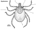
 Hyalomma tick female dorsal.jpg 980 × 1,007; 212 കെ.ബി.
-
 Hyalomma-anatolicum-female-male.jpg 1,229 × 663; 202 കെ.ബി.
-
 Hyalomma-female-dorsal.png 4,126 × 3,369; 196 കെ.ബി.
-
 Hyalomma-male-dorsal-ventral.png 4,399 × 2,759; 271 കെ.ബി.
-
 Hyalomma-rufipes-female-male.jpg 1,225 × 597; 191 കെ.ബി.
-
 IMGP7610-Hyalella azteca with acanthocephalan in body cavity!.jpg 1,563 × 870; 756 കെ.ബി.
-
 Ixodes-female-dorsal-ventral.png 4,654 × 2,993; 244 കെ.ബി.
-
 Ixodes-holocyclus-female-male.jpg 1,178 × 585; 193 കെ.ബി.
-
 Ixodes-male-dorsal-ventral.png 4,534 × 2,214; 172 കെ.ബി.
-
 Leishmania (02).jpg 1,024 × 768; 183 കെ.ബി.
-
 Leishmania (03).jpg 1,024 × 768; 227 കെ.ബി.
-
 Leishmania (04).jpg 491 × 337; 25 കെ.ബി.
-
 Leishmania (05).jpg 946 × 946; 94 കെ.ബി.
-
 Leishmania (06).tif 527 × 544; 434 കെ.ബി.
-
 Leishmania (07).tif 790 × 756; 780 കെ.ബി.
-
 Leishmania (08).tif 715 × 707; 487 കെ.ബി.
-
 Leishmania (09).tif 874 × 662; 726 കെ.ബി.
-
 Leishmania amastigote.png 1,617 × 2,099; 584 കെ.ബി.
-
 Leishmania amastigotes (02).jpg 1,024 × 768; 238 കെ.ബി.
-
 Leishmania brasiliensis (01).tif 2,940 × 1,980; 6.95 എം.ബി.
-
 Leishmania donovani (01).tif 2,944 × 1,976; 6.26 എം.ബി.
-
 Leishmania donovani (02).jpg 1,552 × 1,584; 307 കെ.ബി.
-
 Leishmania infantum (01).jpg 3,264 × 2,448; 1.44 എം.ബി.
-
Leishmania major promastigotes (01).ogv 5.3സെ, 1,228 × 720; 811 കെ.ബി.
-
 Leishmania promastigote.png 2,360 × 3,020; 614 കെ.ബി.
-
 Leishmania promastigotes (03).tiff 1,813 × 1,206; 4.98 എം.ബി.
-
 Leishmania sp. protozoan (01).tif 1,813 × 1,202; 4.44 എം.ബി.
-
 Leishmania sp. protozoan.tif 1,819 × 1,204; 5.22 എം.ബി.
-
 Leishmania spp. - amastigota 02.jpg 960 × 756; 318 കെ.ബി.
-
 Leishmania spp. - promastigote.jpg 429 × 698; 109 കെ.ബി.
-
 Leishmania spp. - spheromastigote.jpg 440 × 480; 115 കെ.ബി.
-
 Leishmania tropica amastigotes (01).tif 397 × 420; 272 കെ.ബി.
-
Leishmaniamajorpromastigotes.ogv 10സെ, 998 × 720; 2.39 എം.ബി.
-
 Leishmaniasis in a dog.tif 1,805 × 1,200; 5.09 എം.ബി.
-
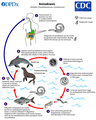
 Leishmaniasis life cycle cdc.tif 3,150 × 2,400; 21.65 എം.ബി.
-
 Lepiselaga-adult-lateral.png 3,640 × 3,336; 221 കെ.ബി.
-
 Lepiselaga-adult.tif 3,640 × 3,336; 200 കെ.ബി.
-
 Life-cycle-of-ixodid-tick.jpg 1,038 × 650; 951 കെ.ബി.
-
 Light microscope photography 9.jpg 1,772 × 1,184; 1.49 എം.ബി.
-
 Linognathus louse female ventral.jpg 1,960 × 1,401; 709 കെ.ബി.
-
 Margaropus-female-dorsal.png 3,666 × 3,275; 198 കെ.ബി.
-
 Margaropus-male-dorsal.png 2,988 × 1,831; 60 കെ.ബി.
-
 McMaster lugemiskamber.jpg 1,632 × 1,224; 99 കെ.ബി.
-
 Megninia-species-mite.jpg 1,055 × 1,581; 699 കെ.ബി.
-
 Morfológica da Giardia muris.jpg 1,182 × 1,280; 145 കെ.ബി.
-
 Myobia-musculi-rodent-mite.jpg 1,196 × 1,814; 869 കെ.ബി.
-
 Neotrombicula larval mite.jpg 1,732 × 1,942; 3.67 എം.ബി.
-
 Numerous serological tests based on immunochromatography in Clinical Microbiology after assaying.jpg 4,160 × 2,340; 3.32 എം.ബി.
-
 Ongoing-challenges-in-the-management-of-malaria-1475-2875-8-S1-S2-1.jpg 600 × 552; 33 കെ.ബി.
-
 Ornithodoros adult soft-tick.jpg 992 × 1,055; 815 കെ.ബി.
-
 Ornithodoros-savignyi-dorsal.jpg 1,865 × 2,176; 778 കെ.ബി.
-
 Otobius-megnini-argasid-tick-nymph.jpg 1,509 × 1,750; 427 കെ.ബി.
-
 Otodectes male female revised.png 5,242 × 3,605; 283 കെ.ബി.
-
 Otodectes-mite-copy.png 5,242 × 3,605; 283 കെ.ബി.
-
 Otodectes-spp-mite.jpg 1,245 × 1,467; 398 കെ.ബി.
-
 Paragonimiasis lifecycle.png 1,300 × 1,308; 868 കെ.ബി.
-
 Parasite (35361000821).jpg 1,149 × 1,578; 257 കെ.ബി.
-

-
 Parásito (Parasite) (35795300224).jpg 4,596 × 7,060; 3.96 എം.ബി.
-
 Parásito de perca, nematode Camallanus corderoi.jpg 1,800 × 2,886; 2.05 എം.ബി.
-
 Parásito digeneo Allocreadium patagonicum.jpg 3,729 × 5,639; 1.39 എം.ബി.
-
 Parásito digeneo Homalometron papilliferum.jpg 3,783 × 5,781; 2.08 എം.ബി.
-
 Parásito monogeneno Acolpenteron australes.jpg 3,750 × 5,415; 2.17 എം.ബി.
-
 Parásito monogeneo Dactylogyrus extensus.jpg 4,161 × 5,646; 2.75 എം.ബി.
-
 Parásito monogeneo Duplaccessorius andinus.jpg 3,918 × 5,341; 3.92 എം.ബി.
-
 Parásito monogeneo Gyrodactylus sp.jpg 3,271 × 5,098; 2.94 എം.ബി.
-
 Parásito nematode Camallanus corderoi.jpg 1,456 × 2,457; 1.19 എം.ബി.
-
 Parásito Steganoderma szidati.jpg 3,702 × 5,666; 1.38 എം.ബി.
-
 Parásito trematode Derogenes lacustris.jpg 2,542 × 4,491; 1.89 എം.ബി.
-
 Parásito trematodes Acanthostomoides apophalliformis en campo oscuro.jpg 3,830 × 5,543; 3.33 എം.ബി.
-
 Parásito trematodes Acanthostomoides apophalliformis.jpg 4,302 × 5,526; 3.92 എം.ബി.
-
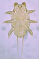
 Pediculus humanus adult (01).png 2,236 × 3,247; 1.16 എം.ബി.
-
 Pediculus humanus egg (01).png 1,419 × 2,635; 349 കെ.ബി.
-
 Phlebotomus (01).png 4,779 × 3,505; 1.6 എം.ബി.
-
 Phormia-larva-adult.png 4,748 × 2,920; 249 കെ.ബി.
-
 Plamodium paludisme (02).png 1,371 × 1,524; 239 കെ.ബി.
-
 Plasmodium female gamete.png 919 × 1,126; 139 കെ.ബി.
-
 Plasmodium merozoites.png 810 × 1,244; 113 കെ.ബി.
-
 Plasmodium oocyste (01.png 863 × 923; 109 കെ.ബി.
-
 Plasmodium paludism.png 1,359 × 737; 164 കെ.ബി.
-
 Plasmodium sporozoites.png 2,100 × 1,021; 298 കെ.ബി.
-
 Plasmodium zygote (01).png 832 × 842; 123 കെ.ബി.
-
 Protozoan parasites and their viral endosymbionts.webp 1,773 × 2,470; 363 കെ.ബി.
-
 Psoroptes-cuniculi-ear-canker-mite.jpg 1,215 × 1,498; 532 കെ.ബി.
-
 Pthirus inguinalis adult.png 3,027 × 3,041; 1.48 എം.ബി.
-
 Reduviidae (01).png 4,547 × 3,094; 1.32 എം.ബി.
-
 Rhipicephalus-appendiculatus-female-male-dorsal.jpg 1,206 × 576; 256 കെ.ബി.
-
 Rhipicephalus-appendiculatus-nymphs-cattle-resistance.jpg 1,856 × 1,763; 1.92 എം.ബി.
-
 Rhipicephalus-australis-female-male.jpg 1,260 × 726; 171 കെ.ബി.
-
 Rhipicephalus-evertsi-male-dorsal.jpg 1,201 × 1,280; 399 കെ.ബി.
-
 Rhipicephalus-female-dorsal.png 3,519 × 3,696; 193 കെ.ബി.
-
 Rhipicephalus-male-dorsal-ventral.png 3,765 × 2,536; 218 കെ.ബി.
-
 Rhipicephalus-microplus-ixodid-female-male.jpg 1,201 × 574; 162 കെ.ബി.
-
 Rhipicephalus-pulchellus-female-male.jpg 1,034 × 580; 333 കെ.ബി.
-
 Rhipicephalus-pulchellus-female.jpg 549 × 455; 148 കെ.ബി.
-
 Rhipicephalus-pulchellus-male.jpg 466 × 469; 137 കെ.ബി.
-
 Rhipicephalus-sanguineus-female-male.jpg 1,216 × 724; 353 കെ.ബി.
-
 Rodent from the Sandhills of west central Nebraska.jpg 800 × 533; 182 കെ.ബി.
-
 Sarcophaga-larva-adult-revised.png 5,338 × 4,888; 355 കെ.ബി.
-
 Sarcoptes scabiei (01).png 3,050 × 3,261; 1.11 എം.ബി.
-
 Schistosoma mansoni cercaria.png 2,220 × 1,750; 296 കെ.ബി.
-
 Schistosoma mansoni egg (01).png 1,270 × 1,096; 165 കെ.ബി.
-
 Schistosoma mansoni female.png 2,654 × 3,207; 847 കെ.ബി.
-
 Schistosoma mansoni miracidium (01).png 1,245 × 1,143; 309 കെ.ബി.
-
 Schistosomiasis life cycle.png 2,803 × 2,393; 825 കെ.ബി.
-
 Schistosomiasis Lifecycle.png 538 × 720; 608 കെ.ബി.
-
 Stain and Reagents for Clinical Parasitology Laboratory.jpg 3,264 × 2,448; 2.17 എം.ബി.
-
 Strongyloides Storcoralis Lifecycle Diagram.jpg 2,000 × 1,676; 608 കെ.ബി.
-
 Symphoromyia-Adult-Snipe-fly.png 4,608 × 2,248; 180 കെ.ബി.
-
 Symphoromyia-Snipe-fly-adult.tif 4,608 × 2,248; 168 കെ.ബി.
-
 T.saginata-egg.jpg 1,200 × 900; 389 കെ.ബി.
-
 Taenia cysticercus (01).png 861 × 1,119; 152 കെ.ബി.
-
 Taenia egg (01).png 906 × 1,042; 219 കെ.ബി.
-
 Taenia saginata (01).png 1,999 × 3,463; 877 കെ.ബി.
-
 Taenia solium (01).png 1,965 × 3,449; 868 കെ.ബി.
-
 Tamier parasité 1.jpg 3,096 × 4,128; 3.89 എം.ബി.
-
 Tamier parasité 2.jpg 3,096 × 4,128; 7.94 എം.ബി.
-
 Tamier parasité 3.jpg 3,096 × 4,128; 4.59 എം.ബി.
-
 Tamier parasité 4.jpg 3,456 × 4,608; 6.42 എം.ബി.
-
 Tamier parasité 5.jpg 3,456 × 4,608; 8.42 എം.ബി.
-
 Tamier parasité 6.jpg 4,608 × 3,456; 5.93 എം.ബി.
-
 The life cycle of the wasp M. demolitor and its bracovirus.png 3,300 × 1,434; 2.93 എം.ബി.
-
 Thelazia in cat 2.jpg 4,032 × 3,024; 1.93 എം.ബി.
-
 Thelazia in cat.jpg 4,032 × 3,024; 6.14 എം.ബി.
-
 Title-Figure.tif 4,757 × 1,885; 137 കെ.ബി.
-
 Title-Figure2.tif 4,757 × 1,885; 145 കെ.ബി.
-
 Title-for-Book.png 4,757 × 1,885; 137 കെ.ബി.
-
 ToRCH IgM Test Result.jpg 4,160 × 2,340; 2.25 എം.ബി.
-
 Toxoplasma gondii bradyzoite.png 917 × 971; 95 കെ.ബി.
-
 Toxoplasma gondii gamete.png 1,204 × 789; 137 കെ.ബി.
-
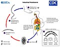
 Toxoplasma gondii Life cycle PHIL 3421 lores hy.png 2,400 × 3,150; 340 കെ.ബി.
-
 Toxoplasma gondii merozoite.png 760 × 1,017; 60 കെ.ബി.
-
 Toxoplasma gondii oocyst spore.png 1,513 × 1,613; 530 കെ.ബി.
-
 Toxoplasma gondii oocyst.png 1,118 × 1,157; 188 കെ.ബി.
-
 Toxoplasma gondii sporocyst.png 979 × 1,176; 288 കെ.ബി.
-
 Toxoplasma LifeCycle CDC.gif 1,130 × 914; 100 കെ.ബി.
-
 Triatoma kissingbug dorsal.jpg 949 × 768; 139 കെ.ബി.
-
 Trichinella spiralis female.png 1,516 × 3,463; 701 കെ.ബി.
-
 Trichinella spiralis larva.png 1,170 × 932; 253 കെ.ബി.


_(19130575244).jpg)


_(20367109545).jpg)


.png)


.jpg)
.jpg)
.jpg)
.jpg)

.jpg)
.jpg)
.jpg)


.jpg)

_(35795300224).jpg)


.png)
.png)
.png)

.png)


.png)


.png)


.png)

.png)

.png)


.png)
.png)
.png)
.png)


