Category:Pus
Zur Navigation springen
Zur Suche springen
gelbliches Exsudat, das im Rahmen einer zellulären Entzündungsreaktion im Körper von Wirbeltieren entsteht  hnisavý zánět spojivek | |||||
| Medium hochladen | |||||
| Audiodatei eines gesprochenen Texts | |||||
|---|---|---|---|---|---|
| Ist ein(e) |
| ||||
| Unterklasse von |
| ||||
| |||||
Unterkategorien
Es werden 2 von insgesamt 2 Unterkategorien in dieser Kategorie angezeigt:
In Klammern die Anzahl der enthaltenen Kategorien (K), Seiten (S), Dateien (D)
A
- Pus in animals (8 D)
H
- Hypopyon (3 D)
Medien in der Kategorie „Pus“
Folgende 27 Dateien sind in dieser Kategorie, von 27 insgesamt.
-
 A Course of Shingles diagram.svg 488 × 477; 332 KB
-
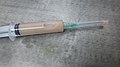 A full of syringe having wound drainage.jpg 4.000 × 2.250; 2,91 MB
-
 Abszess.jpg 416 × 386; 20 KB
-
 Bacterial infection in cuticle.jpg 1.600 × 1.200; 465 KB
-
 Brachial Fistula.JPG 3.072 × 2.304; 2,54 MB
-
 Cholangitis.jpg 420 × 428; 57 KB
-
 Culture swab with blood.jpg 1.600 × 1.200; 513 KB
-
 Fungal UTI picture of urine microscopy showing plenty of yeast cells and pus cells.jpg 3.264 × 2.448; 896 KB
-
 Gram Negative Rods and Pus cells in Gram staining.jpg 4.000 × 2.250; 1.002 KB
-
 Gram positive cocci in chains.jpg 4.000 × 2.250; 2,09 MB
-
 Ideal smear of Sputum.jpg 4.000 × 3.000; 2,64 MB
-
 Non-Ideal smear of Sputum.jpg 4.000 × 3.000; 2,3 MB
-
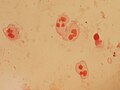
 Normal flora, Pus cells and Epithelial cells.jpg 4.000 × 3.000; 1,39 MB
-
 Numerous Gram Negative Bacteria and Pus cells in Gram staining of sputum.jpg 4.000 × 2.250; 1.020 KB
-
 Numerous pus cells and yeast cells in urine.jpg 3.264 × 2.448; 1,17 MB
-
 Packed pus cells and bacteria in urine sediment microscopy.jpg 3.264 × 2.448; 1,24 MB
-

-
 Plenty of pus cells in Urine Microscopy.jpg 4.000 × 2.250; 2,42 MB
-
 Pleural fluid sediment Microscopy.jpg 8.000 × 6.000; 7,09 MB
-
 Pus cells (dead leukocytes) in urine microscopy.jpg 3.264 × 2.448; 1,02 MB
-
 Pus cells and RBCs in Methylene blue wet mount of pleural fluid.jpg 4.000 × 2.250; 1,68 MB
-
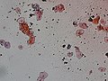
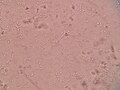
 Pus cells.jpg 4.000 × 3.000; 563 KB
-
 Pus.png 1.520 × 1.225; 1,45 MB
-
 Sputum in Gram stain showing plenty of pus cells.jpg 4.000 × 2.250; 2,24 MB
-
 Streptococcus pyogenes 01 thumbnail.png 240 × 235; 45 KB
-
 Streptococcus pyogenes 01.jpg 2.079 × 2.040; 1,03 MB
-
 Swollen eye with conjunctivitis.jpg 1.407 × 908; 321 KB


_in_urine_microscopy.jpg)


