File:Calcarine sulcus fnana-07-00026-g001.png
Jump to navigation
Jump to search
No higher resolution available.
Calcarine_sulcus_fnana-07-00026-g001.png (337 × 376 pixels, file size: 88 KB, MIME type: image/png)
Captions
Captions
Add a one-line explanation of what this file represents
Summary[edit]
| Description |
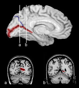
English: Calcarine sulcus (shown in red). Dotted blue line is parieto-occipital sulcus).
|
| Date | |
| Source | Allen JS, Emmorey K, Bruss J and Damasio H (2013) Neuroanatomical differences in visual, motor, and language cortices between congenitally deaf signers, hearing signers, and hearing non-signers. Front. Neuroanat. 7:26. doi:10.3389/fnana.2013.00026 |
| Author | John S. Allen, Karen Emmorey, Joel Bruss and Hanna Damasio. |
Licensing[edit]
This file is licensed under the Creative Commons Attribution 3.0 Unported license.
- You are free:
- to share – to copy, distribute and transmit the work
- to remix – to adapt the work
- Under the following conditions:
- attribution – You must give appropriate credit, provide a link to the license, and indicate if changes were made. You may do so in any reasonable manner, but not in any way that suggests the licensor endorses you or your use.
File history
Click on a date/time to view the file as it appeared at that time.
| Date/Time | Thumbnail | Dimensions | User | Comment | |
|---|---|---|---|---|---|
| current | 07:33, 4 May 2014 | | 337 × 376 (88 KB) | Was a bee (talk | contribs) | {{Information |Description={{en|1=Calcarine sulcus|Calcarine sulcus (shown in red). Dotted blue line is Parieto-occipital sulcus|parieto-occipital sulcus). *'''Top''': Medial surface of left cerebral hemisphere. *'''Bottom''': Coronal... |
You cannot overwrite this file.
File usage on Commons
There are no pages that use this file.