File:Giant anteater at MAV-USP.jpg
Jump to navigation
Jump to search

Size of this preview: 800 × 450 pixels. Other resolutions: 320 × 180 pixels | 640 × 360 pixels | 1,024 × 576 pixels | 1,280 × 720 pixels | 2,560 × 1,440 pixels | 4,241 × 2,386 pixels.
Original file (4,241 × 2,386 pixels, file size: 1.35 MB, MIME type: image/jpeg)
Captions
Captions
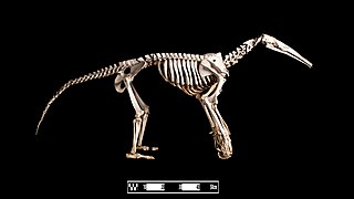
Giant anteater. Myrmecophaga tridactyla”. Specimen of giant anteater skeleton prepared by the bone maceration technique and on display at the Museum of Veterinary Anatomy, FMVZ USP.
Summary[edit]
| Description |
العربية: آكل نمل عملاق. Myrmecophaga tridactyla. نموذج من هيكلٍ عظميّ حُضِّر بتقنية نقع العظام وهو معروضٌ في متحف التشريح الطبي التابع لكلية العلوم الحيوانيَّة والبيطرة في المكسيك، تقنية نقع العظام.
English: Giant anteater. Myrmecophaga tridactyla”. Specimen of giant anteater skeleton prepared by the bone maceration technique and on display at the Museum of Veterinary Anatomy, FMVZ USP. |
| Date | |
| Source | Museum of Veterinary Anatomy FMVZ USP |
| Author | Museum of Veterinary Anatomy FMVZ USP / Wagner Souza e Silva |
Licensing[edit]
This media was produced by the Museum of Veterinary Anatomy (FMVZ USP) and was licensed as Creative Commons BY-SA 4.0. The MAV is an organ of integration of the School of Veterinary Medicine and Animal Science, University of São Paulo.
MAV-FMVZ USP asks to be cited as shown below. If the photographer name is mentioned, please, cite it after the museum's name. If not, just provide the reference to the museum. Attribution in English: Museum of Veterinary Anatomy FMVZ USP / name of the photographer when stated Attribution in Portuguese: Museu de Anatomia Veterinária da FMVZ USP / nome do fotógrafo quando atribuído |
This file is licensed under the Creative Commons Attribution-Share Alike 4.0 International license.
Attribution: Museum of Veterinary Anatomy FMVZ USP / name of the photographer when stated
- You are free:
- to share – to copy, distribute and transmit the work
- to remix – to adapt the work
- Under the following conditions:
- attribution – You must give appropriate credit, provide a link to the license, and indicate if changes were made. You may do so in any reasonable manner, but not in any way that suggests the licensor endorses you or your use.
- share alike – If you remix, transform, or build upon the material, you must distribute your contributions under the same or compatible license as the original.

|
This image has been assessed under the valued image criteria and is considered the most valued image on Commons within the scope: Myrmecophaga tridactyla skeletons. You can see its nomination here. |
File history
Click on a date/time to view the file as it appeared at that time.
| Date/Time | Thumbnail | Dimensions | User | Comment | |
|---|---|---|---|---|---|
| current | 23:54, 15 March 2019 | | 4,241 × 2,386 (1.35 MB) | Rodrigo.Argenton (talk | contribs) | Reframing, background cleaning, contrast improvement, colour improvement. |
| 12:37, 31 August 2018 |  | 3,872 × 2,592 (1.34 MB) | Sturm (talk | contribs) | User created page with UploadWizard |
You cannot overwrite this file.
File usage on Commons
The following 4 pages use this file:
File usage on other wikis
The following other wikis use this file:
- Usage on ar.wikipedia.org
- Usage on arz.wikipedia.org
- Usage on en.wikipedia.org
- Usage on it.wikipedia.org
- Usage on nl.wikipedia.org
- Usage on pt.wikipedia.org
- Usage on ru.wikipedia.org
- Usage on simple.wikipedia.org
- Usage on www.wikidata.org