File:Röntgen rays and electro-therapeutics - with chapters on radium and phototherapy (1910) (14758219185).jpg
From Wikimedia Commons, the free media repository
Jump to navigation Jump to search

Original file(1,706 × 2,176 pixels, file size: 604 KB, MIME type: image/jpeg)

Captions

Captions

Add a one-line explanation of what this file represents

Summary[edit]

Description
English:

Identifier: rntgenrayselectr00kass (find matches)
Title: Röntgen rays and electro-therapeutics : with chapters on radium and phototherapy
Year: 1910 (1910s)
Authors: Kassabian, Mihran Krikor, 1870-1910
Subjects: Electrotherapeutics X-rays Phototherapy Radiology Radiotherapy
Publisher: Philadelphia & London : J.B. Lippincott Company
Contributing Library: Francis A. Countway Library of Medicine
Digitizing Sponsor: Open Knowledge Commons and Harvard Medical School

View Book Page: Book Viewer
About This Book: Catalog Entry
View All Images: All Images From Book
Click here to view book online to see this illustration in context in a browseable online version of this book.

Text Appearing Before Image:
ion: The restricted diaphragmatic movementsmust be regarded as very suspicious of phthisis. This sign, first referredto by Williams, of Boston, has had no theory advanced to explain itsexistence. I will briefly summarize my investigations which gave birthto the theory that an emphysematous condition of the lungs exists inphthisis. Eokitansky and Brehmer noted that lungs too voluminouscoupled with a small heart characterized the phthisical habitus. If thephysician were to depend on percussion dulness as an evidence of earlyphthisis, the affection would never be recognized; lung resonance, notdulness, is the early physical sign of phthisis. The rays are invaluablein the recognition of emphysema ; in this condition, the lungs seem toolarge for the chest, the diaphragm is low and its excursions restricted. Diseases of the Diaphragm. — In spasm^ says Abrams, dia-phragmatic movements are practically suspended on the affected side. ^Journal of the American Medical Association, May 3, 1902.
Text Appearing After Image:
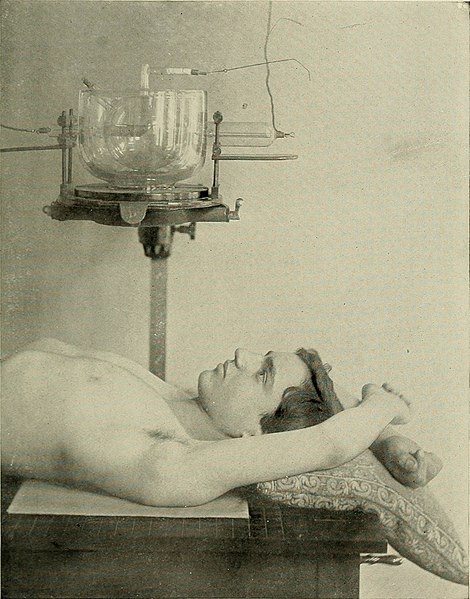
Fig. 176A.—Lungs (dorsal decubitus). Patient lies on a Kin. x 17in. (35 x 42 cm.) plate, with thearms extended over the head : the anode at a distance of 2.5 in. (62 cm.) in the median line, correspond-ing- to the junction of the third and fourth front ribs. This position may be employed in radiographingthe arch and descending aorta. For radiographing the heart, the tube must be displaced downwardto the fifth interspace. APPLICATION OF THE X-EAYS. 305 Suddenly the diapliragm contracts and descends several inches below itsnormal descent. Singultus may accompany the descent, whilst cyanosisand dyspnoea become intense. In paralysis, movements of diaphragmon the affected side are suspended ; during inspiration, the midriff rises.In diaphragmatic pleurisy, movements of the diaphragm are very muchrestricted or even suspended. The upper part of the lung is brighterthan normal, owing to over-distention. ^ Average Xormal Excursion of the Diaphragm.—In quiet breathing,11 centimeters ; betwe

Note About Images

Please note that these images are extracted from scanned page images that may have been digitally enhanced for readability - coloration and appearance of these illustrations may not perfectly resemble the original work.
Date
Source

https://www.flickr.com/photos/internetarchivebookimages/14758219185/

Author Internet Archive Book Images
Permission
(Reusing this file)
At the time of upload, the image license was automatically confirmed using the Flickr API. For more information see Flickr API detail.
Flickr tags
InfoField
  • bookid:rntgenrayselectr00kass
  • bookyear:1910
  • bookdecade:1910
  • bookcentury:1900
  • bookauthor:Kassabian__Mihran_Krikor__1870_1910
  • booksubject:Electrotherapeutics
  • booksubject:X_rays
  • booksubject:Phototherapy
  • booksubject:Radiology
  • booksubject:Radiotherapy
  • bookpublisher:Philadelphia___London___J_B__Lippincott_Company
  • bookcontributor:Francis_A__Countway_Library_of_Medicine
  • booksponsor:Open_Knowledge_Commons_and_Harvard_Medical_School
  • bookleafnumber:442
  • bookcollection:medicalheritagelibrary
  • bookcollection:francisacountwaylibrary
  • bookcollection:americana
Flickr posted date
InfoField
27 July 2014

Licensing[edit]

This image was taken from Flickr's The Commons. The uploading organization may have various reasons for determining that no known copyright restrictions exist, such as:
  1. The copyright is in the public domain because it has expired;
  2. The copyright was injected into the public domain for other reasons, such as failure to adhere to required formalities or conditions;
  3. The institution owns the copyright but is not interested in exercising control; or
  4. The institution has legal rights sufficient to authorize others to use the work without restrictions.

More information can be found at https://flickr.com/commons/usage/.


Please add additional copyright tags to this image if more specific information about copyright status can be determined. See Commons:Licensing for more information.
This image was originally posted to Flickr by Internet Archive Book Images at https://flickr.com/photos/126377022@N07/14758219185. It was reviewed on 16 September 2015 by FlickreviewR and was confirmed to be licensed under the terms of the No known copyright restrictions.

16 September 2015

File history

Click on a date/time to view the file as it appeared at that time.

Date/TimeThumbnailDimensionsUserComment
current04:35, 16 September 2015Thumbnail for version as of 04:35, 16 September 20151,706 × 2,176 (604 KB) (talk | contribs)== {{int:filedesc}} == {{subst:chc}} {{information |description={{en|1=<br> '''Identifier''': rntgenrayselectr00kass ([https://commons.wikimedia.org/w/index.php?title=Special%3ASearch&profile=default&fulltext=Search&search=insource%3A%2Frntgenrayselect...

There are no pages that use this file.