File:Gray708.svg
둘러보기로 이동
검색으로 이동

SVG 파일의 PNG 형식의 미리보기 크기: 550 × 388 픽셀. 다른 해상도: 320 × 226 픽셀 | 640 × 451 픽셀 | 1,024 × 722 픽셀 | 1,280 × 903 픽셀 | 2,560 × 1,806 픽셀
원본 파일 (SVG 파일, 실제 크기 550 × 388 픽셀, 파일 크기: 104 KB)
캡션
설명
이 파일이 나타내는 바에 대한 한 줄 설명을 추가합니다
| 설명 |
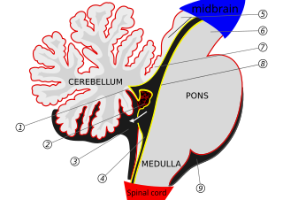
vectorisation of see other version section 1 : posterior medullary velum 2 : Choroid plexus 3 : Cisterna cerebellomedullaris of subarachnoid cavity 4 : Central canal 5 : Corpora quadrigemina 6 : Cerebral peduncle 7 : Anterior medullary velum 8 : Ependymal lining of ventricle 9 : Cisterna pontis of subarachnoid cavityArrow = Flow of cerebrospinal fluid (CSF) through foramen of Magendie |
| 날짜 | |
| 출처 |
 |
| 저자 | lyhana8 |
| 저작권 (이 파일을 인용하기) |
PD |
| 다른 버전 | 이 파일은 다음으로 파생됨: Cerebellum medulla and cortex.svg |
This media file is in the public domain in the United States. This applies to U.S. works where the copyright has expired, often because its first publication occurred prior to January 1, 1929, and if not then due to lack of notice or renewal. See this page for further explanation.
|
 | |
 |
This image might not be in the public domain outside of the United States; this especially applies in the countries and areas that do not apply the rule of the shorter term for US works, such as Canada, Mainland China (not Hong Kong or Macao), Germany, Mexico, and Switzerland. The creator and year of publication are essential information and must be provided. See Wikipedia:Public domain and Wikipedia:Copyrights for more details.
|
파일 역사
날짜/시간 링크를 클릭하면 해당 시간의 파일을 볼 수 있습니다.
| 날짜/시간 | 섬네일 | 크기 | 사용자 | 설명 | |
|---|---|---|---|---|---|
| 현재 | 2021년 10월 17일 (일) 10:43 | | 550 × 388 (104 KB) | Sadopaul (토론 | 기여) | File uploaded using svgtranslate tool (https://svgtranslate.toolforge.org/). Added translation for ko. |
| 2021년 1월 27일 (수) 13:43 |  | 550 × 388 (107 KB) | Pk0001 (토론 | 기여) | up (midbrain) , down(spinal cord) add | |
| 2020년 1월 1일 (수) 17:41 |  | 550 × 388 (106 KB) | SUM1 (토론 | 기여) | Arrow curve disappeared for some reason | |
| 2020년 1월 1일 (수) 17:37 |  | 550 × 388 (106 KB) | SUM1 (토론 | 기여) | Fixed line 8 to actually point to the ependymal lining of ventricle | |
| 2007년 6월 2일 (토) 14:52 |  | 550 × 388 (97 KB) | Edouard-lopez (토론 | 기여) | correction 'Pont' -> 'Pons' | |
| 2006년 11월 25일 (토) 23:51 |  | 550 × 388 (97 KB) | Edouard-lopez (토론 | 기여) | {{Information |Description=vectorisation of Image:Gray708.png 1 : posterior medullary velum 2 : Choroid plexus 3 : Cisterna cerebellodellaris of subarachnoid cavity 4 : Central canal 5 : Corpora quadrigemina 6 : Cerabral peduncle 7 : Anterior medullar |
이 파일을 덮어쓸 수 없습니다.
이 파일을 사용하는 문서
다음 문서 4개가 이 파일을 사용하고 있습니다:
이 파일을 사용하고 있는 모든 위키의 문서 목록
다음 위키에서 이 파일을 사용하고 있습니다:
- ar.wikipedia.org에서 이 파일을 사용하고 있는 문서 목록
- az.wikipedia.org에서 이 파일을 사용하고 있는 문서 목록
- bn.wikipedia.org에서 이 파일을 사용하고 있는 문서 목록
- bs.wikipedia.org에서 이 파일을 사용하고 있는 문서 목록
- cs.wikipedia.org에서 이 파일을 사용하고 있는 문서 목록
- da.wikipedia.org에서 이 파일을 사용하고 있는 문서 목록
- de.wikipedia.org에서 이 파일을 사용하고 있는 문서 목록
- en.wikipedia.org에서 이 파일을 사용하고 있는 문서 목록
- es.wikipedia.org에서 이 파일을 사용하고 있는 문서 목록
- fa.wikipedia.org에서 이 파일을 사용하고 있는 문서 목록
- fr.wikipedia.org에서 이 파일을 사용하고 있는 문서 목록
- gl.wikipedia.org에서 이 파일을 사용하고 있는 문서 목록
- it.wikipedia.org에서 이 파일을 사용하고 있는 문서 목록
- ja.wikipedia.org에서 이 파일을 사용하고 있는 문서 목록
- ko.wikipedia.org에서 이 파일을 사용하고 있는 문서 목록
- mk.wikipedia.org에서 이 파일을 사용하고 있는 문서 목록
- nl.wikipedia.org에서 이 파일을 사용하고 있는 문서 목록
- no.wikipedia.org에서 이 파일을 사용하고 있는 문서 목록
이 파일의 더 많은 사용 내역을 봅니다.