File:HIV entry into T cell schematic.png

원본 파일 (2,805 × 3,405 픽셀, 파일 크기: 4.49 MB, MIME 종류: image/png)
캡션
설명
파일 설명[편집]

|
이 그림 파일은 벡터 SVG 형식으로 다시 만들어야 합니다. SVG의 장점에 대해서는 여기(영어)를 참고해 주십시오. 만약 이 그림의 SVG 버전이 이미 존재한다면 이 틀을 {{vector version available|새 파일 이름.svg}}으로 대체해 주십시오.
|
| 설명 |
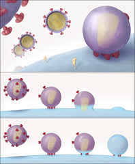
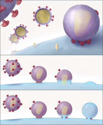
English: "Schematic Representation of the Key Structural Features of SIV and HIV-1 Entry into T Cells"
(A) Different stages of viral entry from budding, to maturation, to entry claw formation. For the SIV strain used here, viruses that are docked to the cell via an entry claw show very few, if any, viral spikes on their surface, whereas non-contacting viruses typically display between 60 and 100 spikes on their surface. The entry claw is composed of between five to seven anchors spanning the region between the virus and the cell, each ∼100 Å long, and spaced laterally by ∼150 Å. (B and C) Two alternative models for viral entry. In the global fusion model (B), the formation of the entry claw is followed by progressive fusion of the viral membrane across its width, leading to merger of the contents of the viral membrane with the cellular membrane. In the local fusion model (C), the formation of the entry claw is followed by the creation of a local pore centered at one of the rods, leading to delivery of the viral core into the cell." |
| 날짜 | Published May 4, 2007 |
| 출처 |
Sougrat R, Bartesaghi A, Lifson JD, et al (May 2007). "Electron tomography of the contact between T cells and SIV/HIV-1: implications for viral entry". PLoS Pathog. 3 (5): e63. PMID 17480119. doi:10.1371/journal.ppat.0030063 Direct link to image: http://www.plospathogens.org/article/showImageLarge.action?uri=info%3Adoi%2F10.1371%2Fjournal.ppat.0030063.g008 |
| 저자 | Rachid Sougrat, Alberto Bartesaghi, Jeffrey D. Lifson, Adam E. Bennett, Julian W. Bess, Daniel J. Zabransky, Sriram Subramaniam |
| 저작권 (이 파일을 인용하기) |
[1] |
| 다른 버전 | JPG version |
라이선스[편집]
|
이 파일은 크리에이티브 커먼즈 저작자표시 2.5 일반 라이선스로 배포됩니다.
|
This file was published in a Public Library of Science journal. Their website states that the content of all PLOS journals is published under the Creative Commons Attribution 4.0 license (or its previous version depending on the publication date), unless indicated otherwise.
|
파일 역사
날짜/시간 링크를 클릭하면 해당 시간의 파일을 볼 수 있습니다.
| 날짜/시간 | 섬네일 | 크기 | 사용자 | 설명 | |
|---|---|---|---|---|---|
| 현재 | 2008년 6월 11일 (수) 00:30 | | 2,805 × 3,405 (4.49 MB) | Fvasconcellos (토론 | 기여) | {{Information |Description="Schematic Representation of the Key Structural Features of SIV and HIV-1 Entry into T Cells" (A) Different stages of viral entry from budding, to maturation, to entry claw formation. For the SIV strain used here, viruses that |
이 파일을 덮어쓸 수 없습니다.
이 파일을 사용하는 문서
다음 문서 2개가 이 파일을 사용하고 있습니다:
이 파일을 사용하고 있는 모든 위키의 문서 목록
다음 위키에서 이 파일을 사용하고 있습니다:
- ar.wikipedia.org에서 이 파일을 사용하고 있는 문서 목록
- en.wikipedia.org에서 이 파일을 사용하고 있는 문서 목록
- es.wikipedia.org에서 이 파일을 사용하고 있는 문서 목록
- ko.wikipedia.org에서 이 파일을 사용하고 있는 문서 목록
- outreach.wikimedia.org에서 이 파일을 사용하고 있는 문서 목록
- sl.wikipedia.org에서 이 파일을 사용하고 있는 문서 목록