Category:Central sulcus
Saltar para a navegação
Saltar para a pesquisa
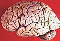
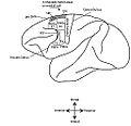
prominent landmark of the brain (primary motor cortex), separating the parietal lobe from the frontal lobe and the primary motor cortex from the primary somatosensory cortex. | |||||
| Carregar ficheiro | |||||
| Instância de |
| ||||
|---|---|---|---|---|---|
| Subclasse de |
| ||||
| Nomeado em referência a | |||||
| Diferente de | |||||
| |||||
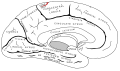
Central sulcus (the fissure of Rolando, the Rolandic fissure).
Multimédia na categoria "Central sulcus"
Esta categoria contém os seguintes 42 ficheiros (de um total de 42).
-
 Blausen 0103 Brain Sensory&Motor ar.png 1 600 × 960; 5,87 MB
-
 Blausen 0103 Brain Sensory&Motor.png 1 600 × 960; 993 kB
-
 Brodmann areas of human lateral frontal cortex.png 1 891 × 889; 2,31 MB
-
 Cambridge Natural History Mammalia Fig 273.png 532 × 374; 33 kB
-
 Central sulcus animation small.gif 150 × 150; 586 kB
-
 Central sulcus animation.gif 300 × 300; 1,75 MB
-
 Central sulcus diagram.png 300 × 190; 27 kB
-
 Central sulcus superior view.png 363 × 366; 134 kB
-
 Central sulcus.png 324 × 209; 27 kB
-
Cerebral Hemisphere Demonstration - Sanjoy Sanyal - Neuroscience Lab Fall 2013 1 (from 1m24s to 2m36s) Central sulcus.webm 1 min 12 s, 852 × 480; 12,71 MB
-
 Cunningham cerebral sulci.png 1 252 × 790; 2,84 MB
-
 Dorsal-cortex front.png 471 × 465; 259 kB
-
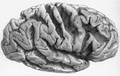
 Einstein brain.jpg 1 022 × 348; 139 kB
-
 Figure3 3-es.png 813 × 615; 358 kB
-
 Figure3 3.jpg 813 × 615; 307 kB
-
 Five sulci measured.png 1 660 × 1 215; 2,36 MB
-
 FrontalCapts.png 2 309 × 911; 927 kB
-
 FrontalCaptsLateral.png 1 252 × 829; 558 kB
-
 Gray1198.png 446 × 350; 24 kB
-
 Gray725 central sulcus.png 255 × 600; 63 kB
-
 Gray726 central sulcus.svg 992 × 573; 143 kB
-
 Gray727 central sulcus.svg 1 025 × 598; 18 kB
-
 Human brain lateral view description.JPG 701 × 487; 49 kB
-
 Human motor cortex topography.png 869 × 551; 32 kB
-

-
 Lateral aspect of left hemispheres showing main sulci.jpg 355 × 860; 275 kB
-
 Lobes.png 909 × 671; 382 kB
-
 LobesCaptsLateral.png 1 201 × 710; 441 kB
-
 LobesCaptsMedial1.png 1 063 × 755; 470 kB
-
 Macaque monkey's premotor areas.jpg 1 200 × 1 108; 156 kB
-
 Motor Cortex monkey.jpg 1 233 × 1 194; 147 kB
-
 ParietCapts lateral.png 1 139 × 758; 407 kB
-
 ParietCapts.png 2 337 × 878; 819 kB
-

-
 Pre- and post-central gyrus, right hemisphere cropped.png 426 × 488; 140 kB
-
 Pre- and post-central gyrus, right hemisphere.jpg 884 × 1 394; 1,13 MB
-
 Sillon occipital ant.png 669 × 519; 122 kB
-
 Sillons vue externe.png 888 × 492; 361 kB
-
 Sulcus centralis - Identification axial - MRI T2.jpg 681 × 795; 69 kB
-
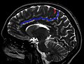 Sulcus centralis - Identification sagittal - MRI T2.jpg 912 × 700; 66 kB
-
 Superficial anatomy of the inferior parietal lobule (IPL).png 454 × 390; 349 kB
-
 Thickness of an humn adult cerebral cortex.jpg 926 × 284; 148 kB


.png)