Category:Diseases and disorders of the eye and adnexa
Перайсці да навігацыі
Перайсці да пошуку
рэнтгеналогія: Ультрагук · камп'ютарная тамаграфія · Magnetic resonance | паталагічная анатомія: Gross pathology · Histopathology | іншы: Fundus · Optical coherence tomography · эпідэміялогія (геаграфічная карта → свет) | фармат файла: SVG · Відэа | 

Categorization according to International Statistical Classification of Diseases and Related Health Problems, 10th Revision.[правіць]
health condition negatively affecting the eye | |||||
| Запампаваць медыя | |||||
| Гэта |
| ||||
|---|---|---|---|---|---|
| Абагульняецца |
| ||||
| |||||
Падкатэгорыі
Паказаны 54 падкатэгорыі з 54.
*
-
A
- Adie syndrome (1 F)
B
- Bitot's spots (2 F)
- Buphthalmos (2 F)
C
- Corectopia (1 F)
- Eye cysts (2 F)
D
F
G
- Globe rupture (3 F)
- Eye gumma (2 F)
H
I
K
- Keratic precipitate (2 F)
L
- Leukocoria (5 F)
N
O
P
S
V
W
- Waardenburg syndrome (18 F)
Старонкі ў катэгорыі «Diseases and disorders of the eye and adnexa»
Паказана 1 старонка гэтай катэгорыі з 1.
Мультымедыя ў катэгорыі "Diseases and disorders of the eye and adnexa"
У гэтай катэгорыі ёсць наступныя 32 файлаў з агульнага ліку 32.
-
 120823-F-CF823-217 (7938218002).jpg 4 256 × 2 832; 6,01 MB
-
 2012-09-22 eye with disease.jpg 2 768 × 2 079; 958 KB
-
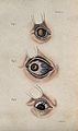 A sheet of three diagrams showing inflamed bloodshot eye def Wellcome V0015923EL.jpg 1 266 × 2 136; 1,53 MB
-
 A sheet of three diagrams showing inflamed bloodshot eye def Wellcome V0015923ER.jpg 1 233 × 2 086; 1,32 MB
-
 A sheet of three diagrams showing inflamed eye defects. Colo Wellcome V0015922EL.jpg 1 272 × 2 082; 1,71 MB
-
 A sheet of three diagrams showing inflamed eye defects. Colo Wellcome V0015922ER.jpg 1 251 × 2 088; 1,49 MB
-
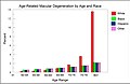 AMD by race and age from National Eye Institute data.jpg 1 882 × 1 216; 289 KB
-

-
 Bilateral222.jpg 666 × 247; 94 KB
-
 Eye discharge.jpg 1 690 × 1 044; 321 KB
-

-

-
-
 Irvine-Gass syndrome .png 1 920 × 1 329; 538 KB
-

-
 Macro Globe oculaire - Mélanome 55-o.apatho-52a-oeil.jpg 639 × 1 499; 500 KB
-
 Macro Globe oculaire - Mélanome 55-o.apatho-52p-oeil.jpg 735 × 540; 195 KB
-
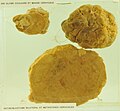 Macro Globe oculaire - Rétinoblastome 55-o.apatho-242a-oeil.jpg 1 676 × 1 547; 1,44 MB
-
 Macro Globe oculaire - Rétinoblastome 55-o.apatho-242p-oeil.jpg 1 563 × 1 497; 1,33 MB
-
 Man with large, malignant growths protruding from both orbits Wellcome L0061863.jpg 4 404 × 5 196; 3,84 MB
-
 Membranous conjunctivitis.jpg 2 023 × 1 125; 570 KB
-
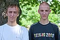
 Nikki Wordsmith Odd Eyes.jpg 828 × 570; 360 KB
-
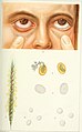
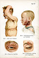
 Pediatrics. (1902) (14577803960).jpg 2 218 × 3 472; 856 KB
-
 PieIXretDiab.jpg 2 322 × 2 224; 923 KB
-
 Reke Salat.png 181 × 216; 50 KB
-
 Retrobulbarbleed.jpg 595 × 552; 102 KB
-
 Siebert 28.jpg 1 945 × 2 866; 1,13 MB
-
 Tape removal.jpg 818 × 615; 110 KB
-
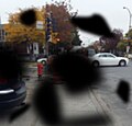
 Vision alterada por SOM diurno.jpg 1 289 × 780; 641 KB
-
 Vision alterada por SOM nocturno.jpg 1 063 × 672; 395 KB
-
 Yamai no Soshi - Eye Disease (part 1).jpeg 2 420 × 2 368; 3,19 MB
-
 Ülekoormuse põhjustatud paistetus.jpg 2 859 × 1 906; 3,3 MB
.jpg)


_(14596082218).jpg)

.png)
_Hrsg._von_W._Kolle_und_A._von_Wassermann_(1912-13)_(16662993341).jpg)


_(14577803960).jpg)


.jpeg)
