Category:Muscles of the human torso
Перайсці да навігацыі
Перайсці да пошуку
| Запампаваць медыя | |||||
| Абагульняецца | |||||
|---|---|---|---|---|---|
| |||||
Падкатэгорыі
Паказаны 19 падкатэгорый з 19.
B
- Bulbospongiosus muscle (4 F)
C
- Cremaster muscles (2 F)
D
E
- External intercostal muscles (17 F)
- External oblique muscles (10 F)
H
I
- Ischiocavernosus muscles (3 F)
L
- Levator ani muscles (2 F)
P
- Pelvic floor (16 F)
R
- Rotatores muscles (2 F)
S
- Serratus anterior muscles (29 F)
T
- Transverse abdominal muscles (3 F)
- Transversus thoracis muscles (1 F)
Мультымедыя ў катэгорыі "Muscles of the human torso"
У гэтай катэгорыі ёсць наступныя 88 файлаў з агульнага ліку 88.
-
 'Back of Male Torso' by Thomas Eakins.JPG 1 926 × 3 210; 757 KB
-
 'Front of Male Torso' by Thomas Eakins.JPG 2 095 × 2 825; 714 KB
-
 Gray388.png 500 × 196; 20 KB
-
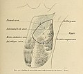
 Gray390.png 391 × 626; 89 KB
-
 Diaphragm - 2.png 600 × 530; 170 KB
-
 Diaphragma.png 600 × 530; 221 KB
-
 Gray391.png 600 × 530; 130 KB
-
 Image391-blank.gif 600 × 530; 165 KB
-
 Gray392 heb.png 692 × 868; 936 KB
-
 Gray392-2.jpg 519 × 650; 412 KB
-
 Gray395.png 483 × 600; 58 KB
-
 Gray396.png 350 × 500; 62 KB
-
 Gray397.png 462 × 700; 63 KB
-
 Gray398.png 550 × 397; 37 KB
-
 Gray398mod.png 550 × 397; 115 KB
-
 Gray399 es.svg 881 × 329; 35 KB
-
 Gray399 esp.jpg 881 × 329; 113 KB
-
 Gray399.png 498 × 186; 11 KB
-
 Gray400.png 503 × 188; 11 KB
-
 Gray401.png 450 × 445; 31 KB
-
 Gray401mod.png 700 × 823; 167 KB
-
 Ligamento Inguinale.jpg 450 × 445; 112 KB
-
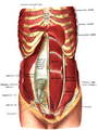
 1112 Muscles of the Abdomen.jpg 1 046 × 1 351; 658 KB
-
 1114 Thorax big.png 1 400 × 1 254; 776 KB
-
 1114 Thorax.jpg 2 275 × 1 273; 927 KB
-
 1119 Muscles that Move the Humerus.jpg 2 229 × 2 371; 1,82 MB
-

-
 A Series of Anatomical Plates Muscles Plate 21.jpg 800 × 1 142; 173 KB
-
 Alphonse Lami; echorche torso, 1861 Wellcome L0025113.jpg 1 136 × 1 734; 793 KB
-
 An écorché figure, trunk, limbs, and head Wellcome V0008849.jpg 2 220 × 3 153; 3,15 MB
-
-
 Mickey Avalon shot by Kris Krug.jpg 5 616 × 3 744; 6,53 MB
-
 Braus 1921 103.png 1 652 × 2 652; 12,56 MB
-
 Braus 1921 105.png 1 540 × 740; 3,27 MB
-
 Braus 1921 107.png 1 600 × 1 652; 7,58 MB
-
 Braus 1921 115.png 1 620 × 1 584; 7,36 MB
-
 Braus 1921 120.png 1 640 × 2 632; 12,37 MB
-
 Braus 1921 85.png 1 180 × 1 048; 3,54 MB
-
 Braus 1921 90.png 1 604 × 2 572; 11,82 MB
-
 Braus 1921 94.png 1 528 × 1 332; 5,83 MB
-
 Braus 1921 95.png 1 648 × 2 692; 12,72 MB
-
 Braus 1921 98.png 1 524 × 1 628; 7,11 MB
-
 Cavité abdominale L4.jpg 726 × 473; 60 KB
-
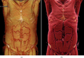
 Contrast-enhanced 3-D CT images of a patient with polytrauma.png 1 302 × 1 041; 1,31 MB
-

-
 Four écorché figures, showing the deep muscles of the axial skeleton Wellcome V0008870.jpg 2 338 × 2 985; 3,04 MB
-
 Gerard de Lairesse - Planche d'anatomie pour Govert Bidloo.jpg 1 200 × 1 991; 412 KB
-
 Grant 1962 11.png 4 432 × 3 816; 13,01 MB
-
 Gray1215 zh.png 463 × 500; 146 KB
-
 Gray1215-ar.png 463 × 500; 95 KB
-
 Gray1215.png 463 × 500; 86 KB
-
 Gray392.png 519 × 650; 75 KB
-
 Grays Anatomy image392.png 519 × 650; 83 KB
-
 Illu trunk muscles zh.jpg 520 × 307; 43 KB
-
 Illu trunk muscles.jpg 520 × 307; 36 KB
-
 J.F. Gautier D'Agoty, Anatomie generale des Wellcome L0021128.jpg 1 220 × 1 578; 1 010 KB
-
 J.F. Gautier D'Agoty, Anatomie generale des Wellcome L0021129.jpg 1 218 × 1 582; 649 KB
-
 Labled diagram of the muscles of the human back.svg 512 × 384; 76 KB
-
 Lord Leighton - The Sluggard back left V&A 2009.jpg 1 962 × 2 874; 772 KB
-
 Mucles de la face posterieure du trone.gif 1 063 × 1 705; 155 KB
-
 Muscles de la face anterieure du trone.gif 1 031 × 1 644; 174 KB
-
 Muscles of the lower limbs and the trunk; two écorché figure Wellcome V0008229.jpg 3 276 × 2 317; 3,62 MB
-
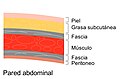 Pared abdominal.jpg 1 792 × 1 180; 290 KB
-
 Plate 62, muscles of the trunk Wellcome L0076997.jpg 5 103 × 7 154; 11,97 MB
-
 Repères anatomiques simples corps antérieur.jpg 515 × 650; 47 KB
-
 Slide9111.JPG 960 × 720; 129 KB
-
 Sobo 1909 237.png 2 132 × 1 232; 7,53 MB
-
 Sobo 1909 245.png 2 308 × 3 300; 21,83 MB
-
 Sobo 1909 246.png 2 160 × 1 644; 10,18 MB
-
 Sobo 1909 247.png 2 036 × 3 352; 19,56 MB
-
 Sobo 1909 248.png 2 400 × 772; 5,31 MB
-
 Sobo 1909 249-esp.jpg 2 388 × 892; 779 KB
-
 Sobo 1909 249.png 2 388 × 892; 6,11 MB
-
 Sobo 1909 250.png 2 208 × 2 944; 18,63 MB
-
 Sobo 1909 252.png 2 428 × 3 208; 22,32 MB
-
 Sobo 1909 253.png 1 740 × 1 848; 9,22 MB
-
 Sobo 1909 254.png 1 770 × 2 076; 10,53 MB
-
 Sternum3.png 533 × 711; 388 KB
-

-
 The breast- its anomalies, its diseases, and their treatment (1917) (14733946156).jpg 1 713 × 1 529; 695 KB
-
 The breast- its anomalies, its diseases, and their treatment (1917) (14753787341).jpg 1 402 × 1 162; 521 KB
-
 The common frog (Page 97, Fig. 60) BHL7743530.jpg 2 099 × 3 308; 556 KB
-
 The common frog (Page 98, Fig. 61) BHL7743529.jpg 2 099 × 3 308; 560 KB
-
 Thorax section 10.jpg 960 × 720; 122 KB
-
 Thorax-diaphragm.png 391 × 626; 85 KB
-
 Vesling "Syntagma...", 1647; male figures Wellcome L0007884.jpg 1 192 × 1 616; 865 KB
-
 Volume Rendering and Cinematic Rendering of a whole-body CT scan.png 1 188 × 822; 1,2 MB
-
 Volume rendering of abdominal muscles.jpg 771 × 943; 219 KB


_(14597836737).jpg)


_(14595709599).jpg)


_(14763390264).jpg)


_(14570314608).jpg)
_(14733946156).jpg)
_(14753787341).jpg)
_BHL7743530.jpg)
_BHL7743529.jpg)


