Category:Porifera
Naviqasiyaya keç
Axtarışa keç
phylum of animals  | |||||||
| Media yüklə | |||||||
| Anlayışın sinfi | |||||||
|---|---|---|---|---|---|---|---|
| Alt sinfidir | |||||||
| Bundan fərqlidir | |||||||
| |||||||
| |||||||
| Takson müəllifi | Robert Edmond Grant, 1836 | ||||||
| |||||||
Wikispecies has an entry on:
- For cleaning and bathing sponges, see category:cleaning sponges.
Alt kateqoriyalar
Bu kateqoriyada 20 altkateqoriya var və onlardan 20 altkateqoriya aşağıda göstərilir.
!
- Quality images of sponges (34 F)
*
?
A
B
- Porifera on black background (23 F)
C
D
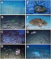
- Deep Sea Abyssal Sponges (144 F)
H
I
†
"Porifera" kateqoriyasındakı fayllar
Cəmi 228 fayldan 200 fayl bu kateqoriyadadır.
(əvvəlki səhifə) (növbəti səhifə)-
 Janssens, Abraham - Lascivia.jpg 836 × 1.000; 811 KB
-

-
 A network of spicules from a leuconoid sponge.jpg 1.280 × 720; 203 KB
-
 A sponge cup with contents at Ponta do Ouro, Mozambique (6661101947).jpg 1.024 × 744; 144 KB
-

-

-
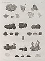
 American journal of pharmacy (1881) (14595707440).jpg 1.822 × 2.620; 2,25 MB
-
 Animals from Coral Patch Seamount (cropped 2).png 362 × 226; 199 KB
-
 Animals from Coral Patch Seamount (cropped 3).png 362 × 225; 174 KB
-
 Animals from Coral Patch Seamount (cropped).png 363 × 225; 197 KB
-
 AquariumRhodesMuseumPorifera.jpg 3.072 × 2.304; 2,54 MB
-
 Armoured sponge.jpg 2.996 × 2.005; 1,05 MB
-
 Asconoid Sponge body plan.png 1.068 × 1.232; 300 KB
-

-
 Sponge Market, Key West Harbor, Fla (NYPL b11707403-G90F127 020F).tiff 3.072 × 1.745; 15,34 MB
-
 Sponge Market, Key West Harbor, Fla (NYPL b11707403-G90F127 020B).tiff 3.072 × 1.778; 15,63 MB
-
 Zoologie. Zoophytes. Éponges charnues, Éponges à piquans (NYPL b14212718-1268613).jpg 4.052 × 5.390; 4,15 MB
-
 Zoologie. Zoophytes. Éponges charnues, Éponges à piquans (NYPL b14212718-1268613).tiff 4.952 × 6.299; 89,26 MB
-
 Zoologie. Zoophytes. Éponges à réseau (NYPL b14212718-1268614).jpg 3.943 × 5.304; 3,91 MB
-
 Zoologie. Zoophytes. Éponges à réseau (NYPL b14212718-1268614).tiff 4.936 × 6.299; 88,98 MB
-
 Zoologie. Zoophytes. Éponges à réseau (NYPL b14212718-1268615).jpg 4.101 × 2.892; 2,1 MB
-
 Zoologie. Zoophytes. Éponges à réseau (NYPL b14212718-1268615).tiff 6.299 × 4.912; 88,54 MB
-
 Barrel Sponge Davy Crocker Reef 20230715.jpg 5.184 × 3.888; 9,95 MB
-
 Black-ball sponge Snapper Ledge 20080310.jpg 2.816 × 2.112; 2,38 MB
-
 Brockhaus and Efron Encyclopedic Dictionary b18 846-0.jpg 873 × 535; 71 KB
-
 Brockhaus and Efron Encyclopedic Dictionary b18 847-1.jpg 679 × 485; 25 KB
-
 Brockhaus and Efron Encyclopedic Dictionary b18 847-2.jpg 1.033 × 939; 97 KB
-

-
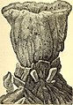
 Cauldron sponge.jpg 3.072 × 2.304; 2,23 MB
-

-

-
 Chelae and sigmancistras in carnivorous sponges.png 1.511 × 1.074; 571 KB
-
 Clathrina coriacea (Montagu, 1818), Diplosoma spongiforme (Giard, 1872).jpg 2.747 × 2.080; 5,25 MB
-

-

-

-
-
 Coral and sponge at Alphard Banks P4100530.jpg 4.000 × 3.000; 2,73 MB
-

-

-

-
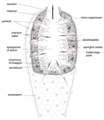 Diagram Bekerspons.png 1.350 × 1.558; 785 KB
-
 Die Gartenlaube (1890) 080.jpg 2.448 × 3.264; 1,71 MB
-
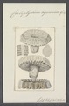
 Die Kalkschwämme. Eine Monographie (1872) (20307658283).jpg 1.404 × 2.326; 834 KB
-
 Diversity of spicule morphology and sets of selected sponge species.jpg 1.200 × 678; 249 KB
-
 Encrusting Sponge (Nara nematifera) (6056494768).jpg 4.416 × 2.649; 4,66 MB
-
 Entobia gallery 041316.jpg 2.496 × 1.696; 2,03 MB
-
 Epitheliozoa collage.png 740 × 1.125; 1,37 MB
-
 Euretidae Gymnorete 1.jpg 1.600 × 1.040; 494 KB
-
 Evolution in the past (Plate 28) BHL21155497.jpg 2.736 × 3.762; 693 KB
-
 Expl6396 (9737645228).jpg 1.920 × 1.080; 1,28 MB
-
 Farreidae Aspidoscopulia.jpg 1.600 × 1.040; 450 KB
-
 Figure 27 03 01.jpg 741 × 286; 115 KB
-
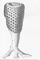 FMIB 49348 Ventriculites simplex, Toulmin Smith.jpeg 622 × 938; 137 KB
-
 FMIB 49349 Ventriculites simplex, Toulmin Smith, Outer surface.jpeg 392 × 379; 76 KB
-

-
 FMIB 49352 Choanites In a fling from the white chalk.jpeg 543 × 868; 127 KB
-

-
 Geodiversidade e biodiversidade do Recife Amazonas.jpg 743 × 846; 852 KB
-
 Global-Diversity-of-Sponges-(Porifera)-pone.0035105.g001.jpg 430 × 496; 233 KB
-
 Global-Diversity-of-Sponges-(Porifera)-pone.0035105.g002.jpg 484 × 561; 230 KB
-
 Global-Diversity-of-Sponges-(Porifera)-pone.0035105.g004.jpg 484 × 344; 95 KB
-
 Grantia Long Section- oscular tip.jpg 2.048 × 1.536; 1,22 MB
-
 Grantia LS 100x.jpg 1.024 × 768; 645 KB
-
 Grantia Spicules.jpg 1.024 × 768; 386 KB
-
 Grey ball sponge at Kreef Reef P5280131.jpg 4.608 × 3.456; 7,33 MB
-
 Halichondria -Hymeniacidon-°grand pont 2225.jpg 3.456 × 2.592; 3,14 MB
-
-
 Highly flexible spicules of marine glass sponge Rossella fibulata.jpg 1.500 × 331; 265 KB
-

-
 Holothuria pyxis.jpg 1.267 × 1.920; 990 KB
-
 Homoscleromopha diversity.png 1.511 × 2.026; 2,92 MB
-
 Journal.pone.0082306.g006.png 2.049 × 2.652; 4,62 MB
-
 Large sponge group (50670094043).jpg 1.920 × 2.560; 6,58 MB
-
 Lecosolenia 100 x.jpg 1.024 × 768; 545 KB
-
 Leucosolenia 400x.jpg 1.024 × 768; 459 KB
-
 Leucosolenia choanocytes.jpg 1.024 × 768; 447 KB
-
 Leucosolenia sections 2.jpg 1.280 × 720; 73 KB
-
 Leucosolenia sections 3.jpg 1.280 × 720; 73 KB
-
 Leucosolenia sections.jpg 1.280 × 720; 98 KB
-
 Leucosolenia Skeleton-1.jpg 1.280 × 720; 55 KB
-
 Leucosolenia Skeleton.jpg 1.280 × 720; 58 KB
-
 Leucosolenia.choanocytes.jpg 1.024 × 768; 466 KB
-
 Long Section of Grantia through canal system.jpg 1.280 × 720; 29 KB
-
 Lotrochota baculifera Réunion.jpg 1.200 × 801; 864 KB
-
 Marine glass sponge Monorhaphis chuni.jpg 250 × 761; 21 KB
-
 Meyers b14 s0682 b1.png 350 × 390; 28 KB
-
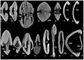
 Morphological diversity of selected marine sponge spicules.jpg 1.046 × 1.200; 160 KB
-

-
 Morphology of sterrasters with smooth rosettes.jpg 1.991 × 2.960; 908 KB
-
 Morphology of sterrasters with warty rosettes.jpg 1.325 × 2.000; 1,49 MB
-
 Observation microscopique d'une éponge.jpg 3.120 × 4.160; 2,82 MB
-
 Osculum (PSF).png 1.319 × 2.964; 510 KB
-

-
 Otavia antiqua 3D reconstruction.jpg 1.300 × 1.000; 618 KB
-
 Photo Sea floor with sea sponges 1959 - Touring Club Italiano 2.2078.jpg 2.164 × 2.200; 500 KB
-
 Photo Sea sponges 1965 - Touring Club Italiano 2.2087.jpg 2.370 × 3.125; 967 KB
-
 Plakortis a IMG 2212.jpg 1.929 × 1.492; 808 KB
-
 Plakortis simplex DSCN3318a.JPG 669 × 659; 150 KB
-
 Plate1proceedingsofgen73zool 0026.jpg 475 × 765; 53 KB
-
 Plate25 Fig 4 journaloflinnean35192224linn 0649 (cropped).jpg 288 × 302; 15 KB
-
 Plate25journaloflinnean35192224linn 0649.jpg 1.757 × 1.298; 284 KB
-
 Plate26 2journaloflinnean35192224linn 0655.jpg 1.688 × 1.331; 286 KB
-
 Plate26 Fig 6a-6e journaloflinnean35192224linn 0655 (cropped).jpg 375 × 402; 20 KB
-
 Plate4proceedingsofgen73zool 0032.jpg 479 × 775; 52 KB
-
 Poribacteria localisation using correlative microscopy.gif 992 × 794; 425 KB
-
 Porifera Arborescent WBRF CEND0313 MP11 STN 134 A1 005.JPG 2.592 × 1.944; 1,76 MB
-
 Porifera Cushion yellow WBRF CEND0313 MP24 STN 152 A1 010.jpg 2.592 × 1.944; 1,54 MB
-
 Porifera encrusting green WBRF CEND0313 MP24 STN 152 A1 010.jpg 2.592 × 1.944; 1,54 MB
-
 Porifera Gemmules 1.jpg 1.280 × 720; 38 KB
-
 Porifera Grantia 8.jpg 1.280 × 720; 63 KB
-
 Porifera Grantia Cross Section 1.jpg 1.280 × 720; 120 KB
-
 Porifera Grantia Cross Section 2.jpg 1.280 × 720; 120 KB
-
 Porifera Grantia Cross Section 3.jpg 1.280 × 720; 91 KB
-
 Porifera Grantia Cross Section 4.jpg 1.280 × 720; 133 KB
-
 Porifera Grantia Long Section Canals.jpg 1.280 × 720; 92 KB
-
 Porifera Grantia Long Section.jpg 1.280 × 720; 92 KB
-
 Porifera Grantia LS.jpg 1.280 × 720; 91 KB
-
 Porifera Grantia Osculum.jpg 1.280 × 720; 58 KB
-
 Porifera Grantia.jpg 1.280 × 720; 75 KB
-
 Porifera grey encrusting WBRF CEND0313 ADDGT43 STN 250 A1 010.jpg 2.592 × 1.944; 1,75 MB
-
 Porifera Lobose orange WBRF CEND0313 ADDGT07 STN 179 A1 007.jpg 2.592 × 1.944; 1,38 MB
-
 Porifera Lobose Yellow WBRF CEND0313 ADDGT06 STN 181 A1 016.jpg 2.592 × 1.944; 1,38 MB
-
 Porifera morphology and internal structure.png 1.345 × 1.553; 3,42 MB
-
 Porifera Orange encrusting WBRF CEND3013 HP12 STN 203 A1 022.JPG 2.592 × 1.944; 1,58 MB
-
 Porifera Pink encrusting WBRF CEND0313 ADDGT43 STN 250 A1 009.jpg 2.592 × 1.944; 1,81 MB
-
 Porifera Red encrusting WBRF CEND0313 ADDGT40 STN 258 A1 017.jpg 2.592 × 1.944; 1,77 MB
-
 Porifera yellow encrusting WBRF CEND0313 ADDGT05 STN 184 A1 033.jpg 2.592 × 1.944; 1,36 MB
-
 Porifera.jpg 4.080 × 3.072; 3,88 MB
-
 Porifera.tif 540 × 540; 504 KB
-
 Presentation and sail of sponges in Symi-2.jpg 4.646 × 2.946; 8,12 MB
-
 Presentation and sail of sponges in Symi-3.jpg 2.108 × 3.044; 4,29 MB
-
 Presentation and sail of sponges in Symi.jpg 4.539 × 2.857; 7,39 MB
-

-

-

-

-

-

-

-

-
 Rod-like collagen-silica-based biomaterial derived in vitro2.jpg 1.306 × 666; 128 KB
-
 Scamp over invertebrates - Grays Reef NMS.jpg 5.616 × 3.744; 11,33 MB
-
 Sclerothamnus D2 Explorer 1.jpg 1.600 × 1.040; 533 KB
-

-

-

-

-
 SEM images of multilayer constructed M. chuni spicule.jpg 600 × 732; 25 KB
-
 Sinoflabrum antiquum.jpg 1.441 × 1.125; 1,6 MB
-
 Sizes of different spicule types of marine sponges.jpg 1.200 × 499; 96 KB
-
 Small coral colony (17053682261).jpg 3.648 × 2.736; 9,13 MB
-
 Spicule formation by sclerocytes in calcareous sponges.jpg 1.961 × 873; 171 KB
-
Spon1000.webm 15 s, 655×480; 3,85 MB
-
 Sponge (PSF).png 1.512 × 1.380; 763 KB
-
 Sponge 01 Molasses Reef 20230714.jpg 5.184 × 3.888; 11,12 MB
-
 Sponge 02 Molasses Reef 20230714.jpg 5.184 × 3.888; 10,87 MB
-
 Sponge 02a Molasses Reef 20230714.jpg 5.184 × 3.888; 10,25 MB
-
 Sponge 03 Molasses Reef 20230714.jpg 5.184 × 3.888; 10,87 MB
-
 Sponge 04 Molasses Reef 20230714.jpg 5.184 × 3.888; 9,19 MB
-
 Sponge 04a Molasses Reef 20230714.jpg 3.888 × 2.916; 6,75 MB
-
 Sponge 05 Molasses Reef 20230714.jpg 5.184 × 3.888; 12,16 MB
-
 Sponge 1 Davy Crocker Reef 20230712.jpg 5.184 × 3.888; 2,97 MB
-
 Sponge 1.jpg 2.592 × 1.936; 1,09 MB
-
 Sponge 2.jpg 2.592 × 1.936; 1,14 MB
-
 Sponge 3.jpg 2.592 × 1.936; 1,11 MB
-
 Sponge at Alphard Banks P4100509.jpg 4.000 × 3.000; 2,57 MB
-
 Sponge at Alphard Banks P4100579.jpg 4.000 × 3.000; 2,44 MB
-
 Sponge at Alphard Banks P4100609.jpg 4.000 × 3.000; 2,58 MB
-
 Sponge at Josh's Reef P5080269.jpg 4.000 × 3.000; 2,58 MB
-
 Sponge at Josh's Reef P5080270.jpg 4.000 × 3.000; 2,49 MB
-
 Sponge at Josh's Reef P5080272.jpg 4.000 × 3.000; 2,48 MB
-
 Sponge at Malindi Reef 1.jpg 4.032 × 3.024; 6,14 MB
-
 Sponge at Malindi Reef 2.jpg 4.032 × 3.024; 6,4 MB
-
 Sponge at Malindi Reef.jpg 4.032 × 3.024; 4,65 MB
-
 Sponge community - Mytilus Seamount.jpg 1.920 × 1.080; 1,43 MB
-
 Sponge consists of a giant basal spicule.jpg 694 × 688; 67 KB
-
 Sponge Davis Reef 20230712.jpg 5.184 × 3.888; 12,59 MB
-
-
 Sponge Gemmules 2.jpg 1.280 × 720; 42 KB
-
 Sponge Gemmules.jpg 1.280 × 720; 42 KB
-
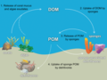 Sponge loop pathway.png 985 × 740; 446 KB
-
 Sponge Market 2017-365-76 (33422830472).jpg 2.416 × 3.541; 1,63 MB
-
 Sponge plazoan nervous system.jpg 920 × 603; 143 KB
-
 Sponge specimen from Natural History Museum Dublin.jpg 3.024 × 4.032; 2,38 MB
-
 Sponge Spicules (3).jpg 1.280 × 720; 198 KB
-
 Sponge Spicules Euplecta (2).jpg 1.280 × 720; 113 KB
-
 Sponge Spicules Euplectella from holdfast region.jpg 1.280 × 720; 48 KB
-
 Sponge Spicules Euplectella.jpg 1.280 × 720; 110 KB
-
 Sponge Spicules of Euplectella.jpg 1.280 × 720; 148 KB
-
 Sponge Spicules.jpg 1.280 × 720; 257 KB
-
 Sponge with basket star at Wilhelm's Wall P3260134.jpg 4.608 × 3.456; 7,38 MB
-
 Sponge.jpg 1.080 × 1.920; 214 KB
-
 Sponge1 (50670921162).jpg 1.920 × 2.560; 4,01 MB
-
 SpongeJI1.jpg 4.117 × 3.088; 1,37 MB
-
 Sponges at 12 Mile Bank P4090160.jpg 4.000 × 3.000; 2,83 MB
-
 Sponges at 12 Mile Bank P4090198.jpg 4.000 × 3.000; 2,71 MB
-
 Sponges at 12 Mile Bank P4090199.jpg 4.000 × 3.000; 2,98 MB
-
 Sponges at 12 Mile Bank P4090240.jpg 4.000 × 3.000; 2,73 MB
-
 Sponges Benwood 20230714.jpg 2.592 × 1.944; 2,99 MB

_(Page_127)_BHL17143698.jpg)

.jpg)
_(14595707440).jpg)
.png)
.png)
.png)


.jpg)
.jpg)
.jpg)


_Front_Cell_Dev_Biol_7_231_fig_9_DE.png)
_Front_Cell_Dev_Biol_7_231_fig_9.jpg)

,_Diplosoma_spongiforme_(Giard,_1872).jpg)
,_Leucosolenia_variabilis_(Haeckel,_1870)_-_Banyuls-sur-Mer_-_07.92.jpg)


_080.jpg)
_(20307658283).jpg)

_(6056494768).jpg)


_BHL21155497.jpg)
.jpg)


.jpeg)

-pone.0035105.g001.jpg)
-pone.0035105.g002.jpg)
-pone.0035105.g004.jpg)


.jpg)


.png)
_From_bodies_of_Porifera,_sea_corals_and_holuthureans.jpg)


.jpg)


.jpg)


_(14744181686).jpg)
_(14764009221).jpg)
_(14787053493).jpg)
_(14787058373).jpg)
_(14579270238).jpg)
_(5988066110).jpg)


.jpg)

.png)


.jpg)


.jpg)


.jpg)
.jpg)


.jpg)


