Category:Protein structural motifs
Jump to navigation
Jump to search
dstinguishing three-dimensional structures characteristically formed by homologous protein sequences | |||||
| Upload media | |||||
| |||||
Subcategories
This category has the following 5 subcategories, out of 5 total.
Media in category "Protein structural motifs"
The following 65 files are in this category, out of 65 total.
-
 1chc animated.gif 640 × 434; 1.23 MB
-
 2m2q.png 500 × 500; 50 KB
-
 310 helix topview.png 378 × 376; 29 KB
-
 Beta Link.pdf 1,239 × 1,752; 588 KB
-
 Beta strand.gif 500 × 167; 749 KB
-
 Classic beta-bulge schematic.png 660 × 860; 163 KB
-
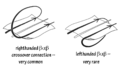 Crossover RH-vs-LH.png 972 × 550; 57 KB
-
 Enlpep5.jpg 237 × 152; 5 KB
-
 Estructura primària i secundària de la proteïna JUNO.png 892 × 374; 134 KB
-
 Estructura secundaria de la rbcL.png 1,026 × 65; 10 KB
-
 Estructura Secundària de l'Esclerostina.png 1,334 × 750; 31 KB
-
 Estructura secundària de la rbcS.png 1,027 × 64; 9 KB
-
 Estructura terciària JUNO.png 502 × 546; 298 KB
-
 Estructura terciària Juno.png 502 × 546; 297 KB
-
 Fipsi.png 369 × 220; 3 KB
-
 Gamma turn of thermolysin.png 2,400 × 2,400; 835 KB
-
 Helice alpha spire 0.png 323 × 658; 40 KB
-
 Helice alpha spire.png 221 × 533; 3 KB
-
 ICK topology1.svg 744 × 1,052; 11 KB
-
 Inverse gamma turn of proteinase A.png 2,400 × 2,400; 1.46 MB
-
 KIX and TAD in complex, structure by Lecoq 2017, PDB 5U4K.jpg 500 × 500; 34 KB
-
 Lasso types.png 834 × 616; 215 KB
-
 LxCxE bound to Retinoblastoma.png 3,257 × 2,202; 1.56 MB
-
 N-terminal of protein.png 365 × 136; 2 KB
-
 Nutlin bound to MDM2.png 2,474 × 2,127; 1.06 MB
-
 PA clan structures 10.png 3,119 × 1,316; 2.43 MB
-
 PA superfamily structural homology.png 7,977 × 7,934; 18.34 MB
-
 Phi.jpg 216 × 232; 7 KB
-
 Pi-helix within an alpha-helix.jpg 1,160 × 2,020; 546 KB
-
 Pyruvate kinase protein domains.png 1,408 × 2,064; 1.37 MB
-
 Ras-P-loop.png 800 × 600; 277 KB
-
 Rossmann fold schematic.svg 1,094 × 354; 177 KB
-
 RpS14.png 840 × 510; 178 KB
-
 RpS15.png 825 × 545; 164 KB
-
 RpS15A.png 560 × 425; 142 KB
-
 RpS16.png 975 × 450; 168 KB
-
 RpS17.png 1,050 × 475; 141 KB
-
 RpS18.png 800 × 550; 187 KB
-
 RpS19.png 750 × 625; 196 KB
-
 RpS2.png 775 × 840; 266 KB
-
 RpS20.png 920 × 350; 113 KB
-
 RpS21.png 750 × 450; 99 KB
-
 RpS23.png 950 × 525; 180 KB
-
 RpS24.png 850 × 550; 173 KB
-
 RpS25.png 650 × 450; 110 KB
-
 RpS26.png 825 × 425; 141 KB
-
 RpS27.png 750 × 325; 106 KB
-
 RpS27A.png 1,075 × 500; 114 KB
-
 RpS28.png 480 × 340; 80 KB
-
 RpS3.png 1,225 × 890; 255 KB
-
 RpS30.png 775 × 425; 90 KB
-
 RpS3A.png 965 × 795; 275 KB
-
 RpS4.png 690 × 845; 276 KB
-
 RpS5.png 815 × 550; 218 KB
-
 RpS6.png 1,650 × 700; 279 KB
-
 RpS7.png 675 × 775; 164 KB
-
 RpS8.png 1,070 × 575; 235 KB
-
 RpS9.png 1,025 × 550; 235 KB
-
 RpSA.png 675 × 580; 215 KB
-
 Schematic C17orf64.png 836 × 308; 35 KB
-
 Sclerostin secondary structure.jpg 1,332 × 254; 26 KB
-
 Structural homology of the PA clan.png 2,021 × 1,982; 2.14 MB
-
 STturn.pdf 1,239 × 1,752; 86 KB
-
 TEV protease beta-barrels.png 510 × 487; 225 KB
-
 UM chem505 1JUN side view.png 820 × 1,575; 525 KB


