File:Gray726.svg

Original file (SVG file, nominally 992 × 573 pixels, file size: 146 KB)
Captions
Captions
Summary[edit]
| Description |
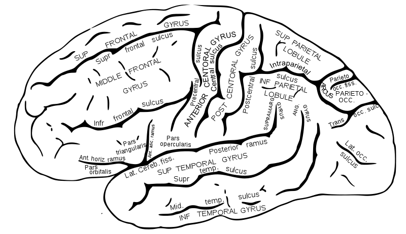
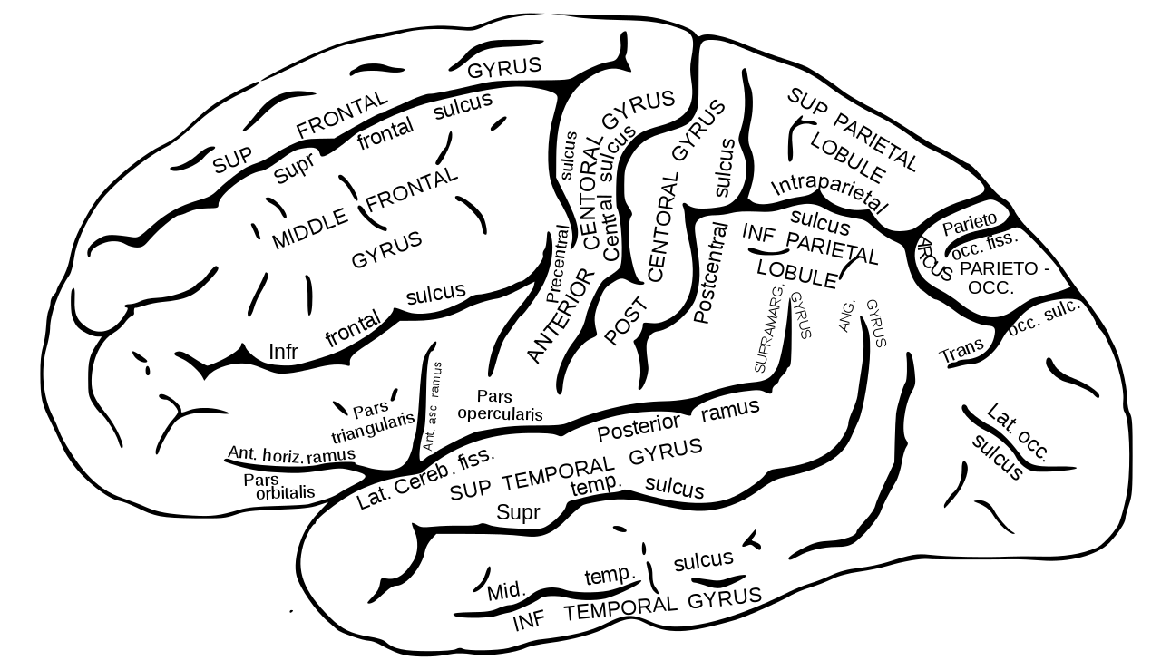
English: Lateral surface of left cerebral hemisphere. |
||||||||||||||||||||
| Plate | 726 | ||||||||||||||||||||
| Date | before 1858 | ||||||||||||||||||||
| Source |
|
||||||||||||||||||||
| Author |
|
||||||||||||||||||||
| Other versions |
  |
||||||||||||||||||||
.jpg)
Book[edit]
| Henry Gray: Gray's Anatomy (20th edition)
|
|||||||||||||||||||||||
|---|---|---|---|---|---|---|---|---|---|---|---|---|---|---|---|---|---|---|---|---|---|---|---|
| Author |
|
-_Title_page.png) | |||||||||||||||||||||
| Editor |
Revised by Warren H. Lewis |
||||||||||||||||||||||
| Illustrator |
|
||||||||||||||||||||||
| Title | |||||||||||||||||||||||
| Edition |
20 |
||||||||||||||||||||||
| Publisher | |||||||||||||||||||||||
| Object type |
version, edition or translation |
||||||||||||||||||||||
| Page overview | list of all the plates | ||||||||||||||||||||||
| Language |
English |
||||||||||||||||||||||
| Publication date |
1918 |
||||||||||||||||||||||
| Place of publication |
Philadelphia / New York City |
||||||||||||||||||||||
| Source | Bartleby | ||||||||||||||||||||||

Licensing[edit]
This image is in the public domain because it is a mere mechanical scan or photocopy of a public domain original, or – from the available evidence – is so similar to such a scan or photocopy that no copyright protection can be expected to arise. The original itself is in the public domain for the following reason:
This tag is designed for use where there may be a need to assert that any enhancements (eg brightness, contrast, colour-matching, sharpening) are in themselves insufficiently creative to generate a new copyright. It can be used where it is unknown whether any enhancements have been made, as well as when the enhancements are clear but insufficient. For known raw unenhanced scans you can use an appropriate {{PD-old}} tag instead. For usage, see Commons:When to use the PD-scan tag.  | ||||
File history
Click on a date/time to view the file as it appeared at that time.
| Date/Time | Thumbnail | Dimensions | User | Comment | |
|---|---|---|---|---|---|
| current | 06:15, 30 October 2009 | | 992 × 573 (146 KB) | Was a bee (talk | contribs) | fix white area |
| 05:47, 28 October 2009 |  | 992 × 573 (145 KB) | Was a bee (talk | contribs) | intraparietal sulcusl -> intraparietal sulcus | |
| 05:35, 28 October 2009 |  | 992 × 573 (141 KB) | Was a bee (talk | contribs) | lat. occ. sulcusl -> lat. occ. sulcus | |
| 13:42, 25 October 2009 |  | 992 × 573 (138 KB) | Was a bee (talk | contribs) | procentral sulcus > precentral sulcus | |
| 03:39, 25 October 2009 |  | 992 × 573 (155 KB) | Was a bee (talk | contribs) | text | |
| 02:47, 25 October 2009 |  | 992 × 573 (155 KB) | Was a bee (talk | contribs) | spelling | |
| 01:56, 25 October 2009 |  | 992 × 573 (519 KB) | Was a bee (talk | contribs) | Path>Object to Path | |
| 01:52, 25 October 2009 |  | 992 × 573 (83 KB) | Was a bee (talk | contribs) | text | |
| 15:18, 24 October 2009 |  | 992 × 573 (117 KB) | Was a bee (talk | contribs) | bug?? | |
| 14:38, 24 October 2009 |  | 992 × 573 (117 KB) | Was a bee (talk | contribs) | Reverted to version as of 14:31, 24 October 2009 |
You cannot overwrite this file.
File usage on Commons
The following 41 pages use this file:
- Gyri
- Sulci (neuroanatomy)
- File:Gray726.png
- File:Gray726.svg
- File:Gray726 Inferior frontal sulcus.svg
- File:Gray726 Superior frontal sulcus.svg
- File:Gray726 angular gyrus.png
- File:Gray726 cant been seen lateral.png
- File:Gray726 central sulcus.svg
- File:Gray726 frontal lobe.png
- File:Gray726 frontal pole.png
- File:Gray726 inferior frontal gyrus.png
- File:Gray726 inferior parietal lobule.png
- File:Gray726 inferior parietal lobule (hy).png
- File:Gray726 inferior temporal gyrus.png
- File:Gray726 intraparietal sulcus.svg
- File:Gray726 lateral occipital gyrus.png
- File:Gray726 lateral sulcus.svg
- File:Gray726 middle frontal gyrus.png
- File:Gray726 middle temporal gyrus.png
- File:Gray726 middle temporal sulcus.svg
- File:Gray726 occipital lobe.png
- File:Gray726 occipital pole.png
- File:Gray726 opecular part of IFG.png
- File:Gray726 orbital part of IFG.png
- File:Gray726 parietal lobe.png
- File:Gray726 parieto-occipital fissure.svg
- File:Gray726 postcentral gyrus.png
- File:Gray726 postcentral sulcus.svg
- File:Gray726 precentral gyrus.png
- File:Gray726 precentral sulcus.svg
- File:Gray726 superior frontal gyrus.png
- File:Gray726 superior parietal lobule.png
- File:Gray726 superior temporal gyrus.png
- File:Gray726 superior temporal sulcus.svg
- File:Gray726 supramarginal gyrus.png
- File:Gray726 temporal lobe.png
- File:Gray726 temporal pole.png
- File:Gray726 trans occipital sulcus.svg
- File:Gray726 triangular part of IFG.png
- File:Gray727 lateral-occipital sulcus.svg
.png)
File usage on other wikis
The following other wikis use this file:
- Usage on de.wikipedia.org
- Usage on en.wikipedia.org
- Usage on en.wikibooks.org
- Usage on fr.wikipedia.org
- Usage on it.wikipedia.org
- Usage on ja.wikipedia.org
- Usage on la.wikipedia.org
- Usage on nl.wikipedia.org