File:Phototransduction.png
Jump to navigation
Jump to search
Size of this preview: 800 × 338 pixels. Other resolutions: 320 × 135 pixels | 640 × 271 pixels | 1,327 × 561 pixels.
Original file (1,327 × 561 pixels, file size: 344 KB, MIME type: image/png)
Captions
Captions
Add a one-line explanation of what this file represents
| Description |
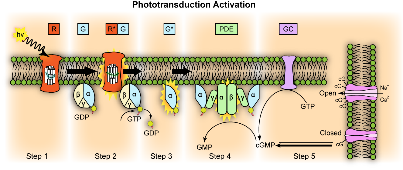
English: Representation of molecular steps in photoactivation (modified from Leskov et al., 2000). Depicted is an outer membrane disk in a rod. Step 1: Incident photon (hv) is absorbed and activates a rhodopsin by conformational change in the disk membrane to R*. Step 2: Next, R* makes repeated contacts with transducin molecules, catalyzing its activation to G* by the release of bound GDP in exchange for cytoplasmic GTP. The α and γ subunits Step 3: G* binds inhibitory γ subunits of the phosphodiesterase (PDE) activating its α and β subunits. Step 4: Activated PDE hydrolyzes cGMP. Step 5: Guanylyl cyclase (GC) synthesizes cGMP, the second messenger in the phototransduction cascade. Reduced levels of cytosolic cGMP cause cyclic nucleotide gated channels to close preventing further influx of Na+ and Ca2+.
Deutsch: Repräsentation der molekularen Schritte der Lichtaktivierung (verändert nach Leskov et al., 2000). Es wird die äussere Membranschiebe eines Stäbchens abgebildet.
|
||
| Date | |||
| Source | http://en.wikipedia.org/wiki/File:Phototransduction.png | ||
| Author | Jason J. Corneveaux, wiki user: Caddymob (talk) | ||
| Permission (Reusing this file) |
I, the copyright holder of this work, hereby publish it under the following licenses:
This file is licensed under the Creative Commons Attribution 3.0 Unported license.
You may select the license of your choice. |
||
| Other versions | Derivative works of this file: Phototransduction uk.png |

File history
Click on a date/time to view the file as it appeared at that time.
| Date/Time | Thumbnail | Dimensions | User | Comment | |
|---|---|---|---|---|---|
| current | 15:42, 18 April 2010 | | 1,327 × 561 (344 KB) | Thomas.haslwanter (talk | contribs) | {{Information |Description=Representation of molecular steps in photoactivation (modified from Leskov et al., 2000). Depicted is an outer membrane disk in a rod. Step 1: Incident photon (hv) is absorbed and activates a rhodopsin by conformational change i |
You cannot overwrite this file.
File usage on Commons
There are no pages that use this file.
File usage on other wikis
The following other wikis use this file:
- Usage on bg.wikibooks.org
- Usage on de.wikibooks.org
- Usage on el.wikibooks.org
- Usage on en.wikipedia.org
- Usage on en.wikibooks.org
- Usage on et.wikipedia.org
- Usage on eu.wikipedia.org
- Usage on fa.wikipedia.org
- Usage on fr.wikibooks.org
- Usage on gl.wikipedia.org
- Usage on it.wikibooks.org
- Usage on ml.wikipedia.org
- Usage on mn.wikipedia.org
- Usage on pt.wikibooks.org