File:1L9H (Bovine Rhodopsin) 2.png
Jump to navigation
Jump to search
Size of this preview: 439 × 600 pixels. Other resolutions: 176 × 240 pixels | 637 × 870 pixels.
Original file (637 × 870 pixels, file size: 322 KB, MIME type: image/png)
Captions
Captions
Add a one-line explanation of what this file represents
Summary[edit]
_2.png&action=edit§ion=1)
| Description |
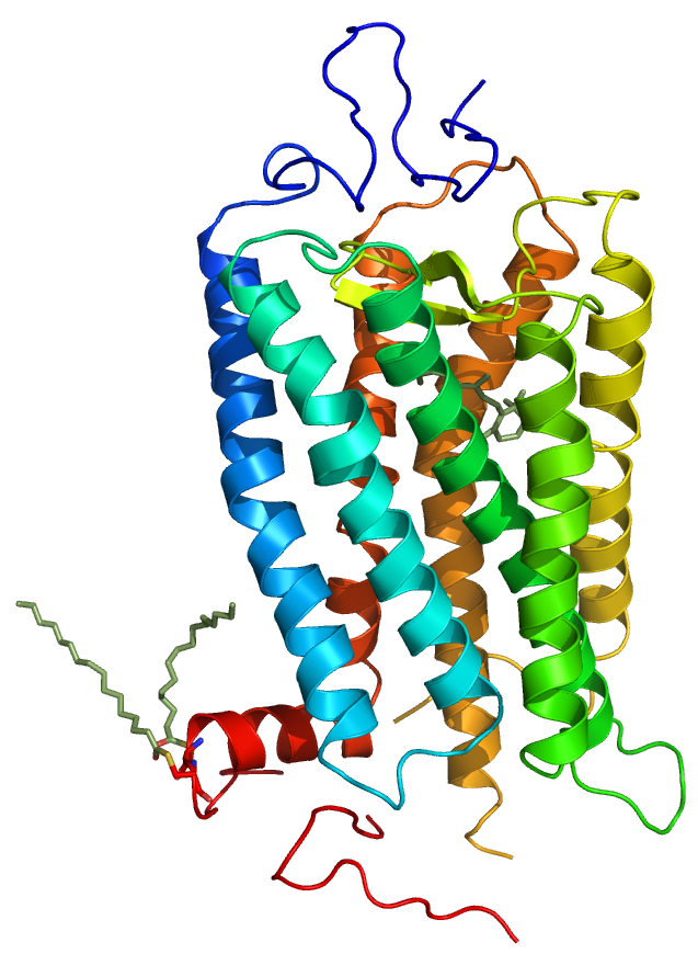
English: 3D structure model of bovine rhodopsin. Derived from the 2.6 Å crystal stucture of rhodopsin (1L9H) with covalently linked retinal and palmityl residues (grey). Structural informations were obtained from pdb.org and rendered using PyMol 0.99. Blue: TMI. Lightblue: TMII. Cyan: TMIII. Green: TMIV. Yellow: TMV. Organge: TMVI. Red-orange: TMVII. Red: Hx8. |
| Date | |
| Source | Own work |
| Author | S. Jähnichen |
Licensing[edit]
_2.png&action=edit§ion=2)
| I, the copyright holder of this work, release this work into the public domain. This applies worldwide. In some countries this may not be legally possible; if so: I grant anyone the right to use this work for any purpose, without any conditions, unless such conditions are required by law. |
File history
Click on a date/time to view the file as it appeared at that time.
| Date/Time | Thumbnail | Dimensions | User | Comment | |
|---|---|---|---|---|---|
| current | 19:41, 20 February 2010 | | 637 × 870 (322 KB) | S. Jähnichen (talk | contribs) | {{Information |Description={{en|1=3D structure model of bovine rhodopsin. Derived from the 2.6 Å crystal stucture of rhodopsin (1L9H) with covalently linked retinal and palmityl residues (grey). Structural informations were obtained from pdb.org and rend |
You cannot overwrite this file.
File usage on Commons
The following page uses this file:
File usage on other wikis
The following other wikis use this file:
- Usage on bs.wikipedia.org
- Usage on de.wikipedia.org
- Usage on es.wikipedia.org
- Usage on pl.wikipedia.org
- Usage on sr.wikipedia.org
_2.png&oldid=748383645)