File:Adenocarcinoma of the prostate.jpg
Jump to navigation
Jump to search

Size of this preview: 800 × 588 pixels. Other resolutions: 320 × 235 pixels | 640 × 470 pixels | 1,024 × 752 pixels | 1,280 × 940 pixels | 2,560 × 1,880 pixels | 4,096 × 3,008 pixels.
Original file (4,096 × 3,008 pixels, file size: 2.27 MB, MIME type: image/jpeg)
Captions
Captions
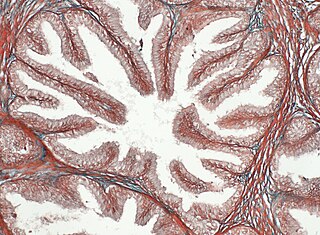
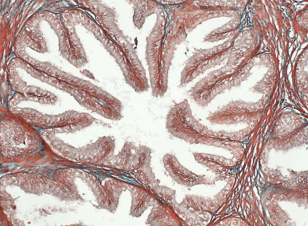
Adenocarcinoma of the prostate
Summary[edit]
| Description |
Українська: На мікрофотографії можна спостерігати м’язово-еластичну строму органу, де гладком’язові елементи зафарбовані червоним кольором, еластичні — зеленим. Кінцеві секреторні відділи передміхурової залози вистелені псевдобагатошаровим епітелієм, який забарвлений у світліший червоний колір. У просвіті видно секрет залози. Зрізи тканини зафарбовані трихромом Масона. Збільшення 200х. Мікрофотографія зроблена в Інституті експериментальної патології, онкології і радіобіології ім. Р.Є. Кавецького НАН України (Київ, Україна); використано мікроскоп Сarl Zeiss Axio Imager A2 з використанням камери Axiocam 712 color. English: Adenocarcinoma of the prostate. On the photomicrograph, you can observe the muscle-elastic stroma of the organ, where the smooth muscle elements are painted in red, and the elastic ones are painted in green. The end secretory parts of the prostate gland are lined with pseudo-multilayered epithelium, which is colored in a lighter red color. The gland secret is visible in the lumen. Fabric sections are stained with Mason's trichrome. Magnification 200x. Photomicrograph taken at the R.E. Kavetsky Institute of Experimental Pathology, Oncology and Radiobiology, National Academy of Sciences of Ukraine (Kyiv, Ukraine); a Carl Zeiss Axio Imager A2 microscope with an Axiocam 712 color camera was used. |
| Date | |
| Source | Own work |
| Author | Endolysosome |
Licensing[edit]
I, the copyright holder of this work, hereby publish it under the following license:
This file is licensed under the Creative Commons Attribution-Share Alike 4.0 International license.
- You are free:
- to share – to copy, distribute and transmit the work
- to remix – to adapt the work
- Under the following conditions:
- attribution – You must give appropriate credit, provide a link to the license, and indicate if changes were made. You may do so in any reasonable manner, but not in any way that suggests the licensor endorses you or your use.
- share alike – If you remix, transform, or build upon the material, you must distribute your contributions under the same or compatible license as the original.
| This image was uploaded as part of Science Photo Competition 2023 in Ukraine. |
File history
Click on a date/time to view the file as it appeared at that time.
| Date/Time | Thumbnail | Dimensions | User | Comment | |
|---|---|---|---|---|---|
| current | 13:45, 20 December 2023 | | 4,096 × 3,008 (2.27 MB) | Endolysosome (talk | contribs) | Uploaded own work with UploadWizard |
You cannot overwrite this file.
File usage on Commons
There are no pages that use this file.