File:Diffuse midline glioma MRI.jpg
Jump to navigation
Jump to search
Size of this preview: 594 × 600 pixels. Other resolutions: 238 × 240 pixels | 475 × 480 pixels | 760 × 768 pixels | 1,014 × 1,024 pixels | 1,794 × 1,812 pixels.
Original file (1,794 × 1,812 pixels, file size: 352 KB, MIME type: image/jpeg)
Captions
Captions
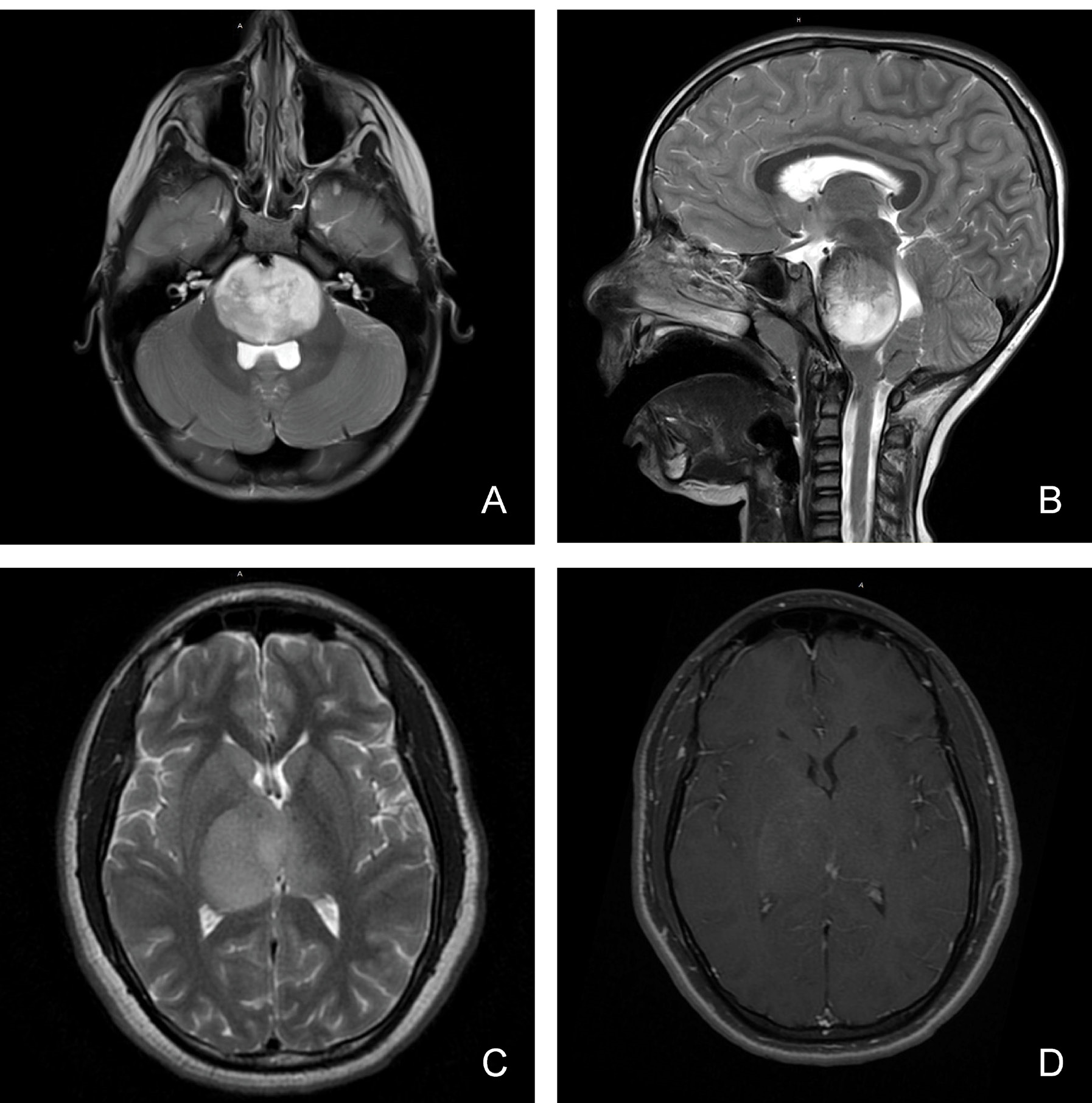
Diffuse midline glioma MRI
Summary[edit]
| Description |
English: Magnetic resonance imaging characteristics (MRI) of pediatric diffuse midline glioma. Pediatric diffuse midline glioma arise in the brainstem (A, B) or thalamus (C, D). T2 weighted MR imaging (A, B, C) demonstrates a homogenous, expansile, infiltrative lesion with extension within white matter tracts. Minimal to heterogenous enhancement may be observed on T1 post-gadolinium sequences (D). Local mass effect and perilesional edema may also be seen. |
| Date | |
| Source | https://doi.org/10.18632/oncotarget.26430 |
| Author | Huang T., Garcia R., Qi J., Lulla R., Horbinski C., Behdad A., Wadhwani N., Shilatifard A., James C., Saratsis A. M. |
| Other versions |
.jpg)
.jpg)
Licensing[edit]
This file is licensed under the Creative Commons Attribution 4.0 International license.
- You are free:
- to share – to copy, distribute and transmit the work
- to remix – to adapt the work
- Under the following conditions:
- attribution – You must give appropriate credit, provide a link to the license, and indicate if changes were made. You may do so in any reasonable manner, but not in any way that suggests the licensor endorses you or your use.
File history
Click on a date/time to view the file as it appeared at that time.
| Date/Time | Thumbnail | Dimensions | User | Comment | |
|---|---|---|---|---|---|
| current | 11:30, 2 December 2023 | | 1,794 × 1,812 (352 KB) | MaligneRange (talk | contribs) | higher res |
| 11:24, 2 December 2023 |  | 675 × 681 (204 KB) | MaligneRange (talk | contribs) | Uploaded a work by Huang T., Garcia R., Qi J., Lulla R., Horbinski C., Behdad A., Wadhwani N., Shilatifard A., James C., Saratsis A. M. from https://doi.org/10.18632/oncotarget.26430 with UploadWizard |
You cannot overwrite this file.
File usage on Commons
The following 2 pages use this file: