File:Dopamine immunoreactive fibres in the prefrontal cortex, nucleus accumbens and amygdala.JPEG
Jump to navigation
Jump to search
Size of this preview: 508 × 599 pixels. Other resolutions: 203 × 240 pixels | 407 × 480 pixels | 651 × 768 pixels | 868 × 1,024 pixels | 2,283 × 2,693 pixels.
Original file (2,283 × 2,693 pixels, file size: 974 KB, MIME type: image/jpeg)
Captions
Captions
Add a one-line explanation of what this file represents
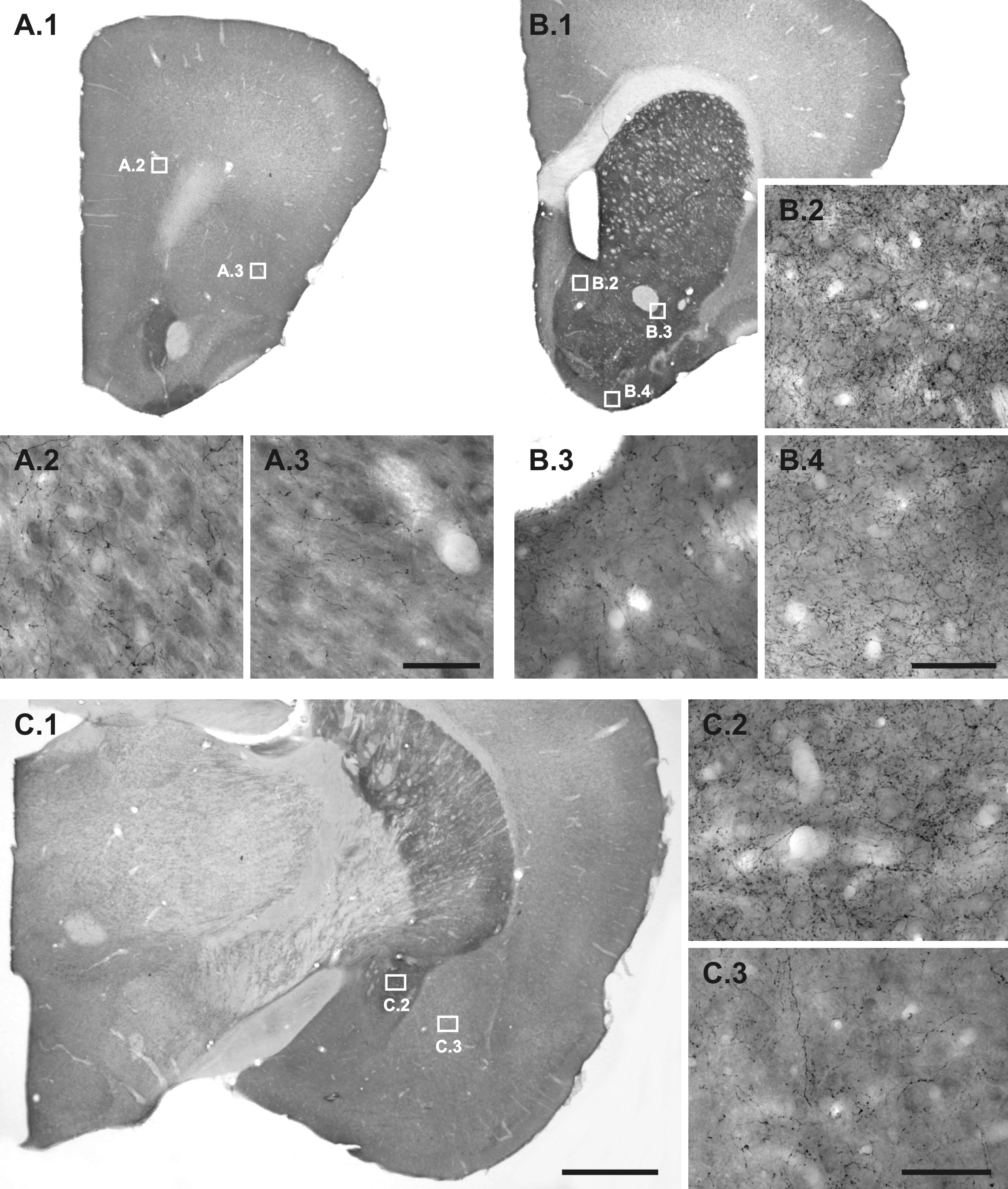
| Description | Comparative image to give an idea of dopaminergic neuron distribution in the prefrontal cortex, nucleus accumbens and amygdala. Figure legend in source article (CC-by-2.0): "Dopamine immunoreactive fibres in each of the quantified regions. Representative photomicrographs, taken from a saline control, of dopamine (DA) immunoreactive fibres of each of the quantified regions. A.1: Prefrontal cortex; A.2: Layer VI of the prelimbic area; A.3: Layer IV of the lateral orbital and agranular insular areas. B.1: Nucleus accumbens (NAC); B.2: Medial shell of NAC; B.3: Lateral core of NAC; B.4: Olfactory tubercle. C.1: Amygdala (AMY); C.2: Central nucleus of AMY; C.3: Basolateral nucleus of AMY. Note the differential innervation pattern and density of DA fibres in the respective regions. Scale bars: 1000 μm (A.1, B.1, C.1); 50 μm (A.2-3, B.2-4, C.2-3)." |
| Date | |
| Source | Brummelte, Susanne; Thorsten Grund, Andrea Czok, Gertraud Teuchert-Noodt, Jorg Neddens (2006). "Long-term effects of a single adult methamphetamine challenge: Minor impact on dopamine fibre density in limbic brain areas of gerbils". Behavioral and Brain Functions 2 (1): 12. DOI:10.1186/1744-9081-2-12. ISSN 1744-9081. Retrieved on 2007-12-21. |
| Author | Article by Susanne Brummelte, Thorsten Grund, Andrea Czok, Gertraud Teuchert-Noodt, Jorg Neddens |
| Permission (Reusing this file) |
This file is licensed under the Creative Commons Attribution 2.0 Generic license.
|
File history
Click on a date/time to view the file as it appeared at that time.
| Date/Time | Thumbnail | Dimensions | User | Comment | |
|---|---|---|---|---|---|
| current | 21:34, 21 December 2007 | | 2,283 × 2,693 (974 KB) | OldakQuill (talk | contribs) | {{Information |Description=Comparative image to give an idea of dopaminergic neuron distribution in the prefrontal cortex, nucleus accumbens and amygdala. Figure legend in source article (CC-by-2.0): "'''Dopamine immunoreactive fibres in each of the quant |
You cannot overwrite this file.
File usage on Commons
There are no pages that use this file.