File:Estructura química de l'ADN.svg
Jump to navigation
Jump to search

Size of this PNG preview of this SVG file: 514 × 600 pixels. Other resolutions: 206 × 240 pixels | 411 × 480 pixels | 658 × 768 pixels | 878 × 1,024 pixels | 1,755 × 2,048 pixels | 1,500 × 1,750 pixels.
Original file (SVG file, nominally 1,500 × 1,750 pixels, file size: 12 KB)
Captions
Captions
Add a one-line explanation of what this file represents
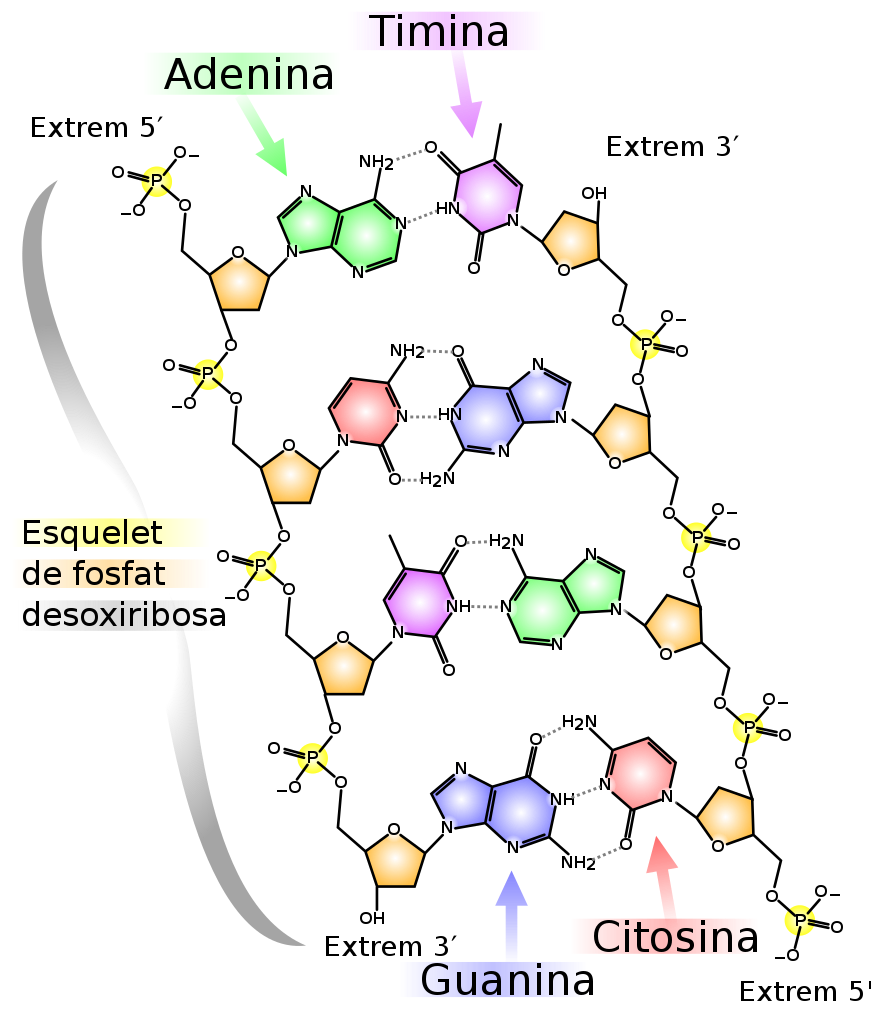
| Description | Estructura derivada de l'ADN, amb etiquetes colorides que identifiquen les quatre bases, així com els components fosfat i desoxiribosa del tronc. Estructura creada amb Chemtool, exportada a SVG, i editada amb Inkscape. Atribuïu la imatge a Madeleine Price Ball si en feu un ús comercial. |
| Date | (UTC) |
| Source | |
| Author |
|
| Other versions |
[]
|


| This is a retouched picture, which means that it has been digitally altered from its original version. Modifications: translated to Catalan. The original can be viewed here: DNA chemical structure.svg:
|
I, the copyright holder of this work, hereby publish it under the following licenses:
This file is licensed under the Creative Commons Attribution-Share Alike 2.5 Generic, 2.0 Generic and 1.0 Generic license.
- You are free:
- to share – to copy, distribute and transmit the work
- to remix – to adapt the work
- Under the following conditions:
- attribution – You must give appropriate credit, provide a link to the license, and indicate if changes were made. You may do so in any reasonable manner, but not in any way that suggests the licensor endorses you or your use.
- share alike – If you remix, transform, or build upon the material, you must distribute your contributions under the same or compatible license as the original.
| This file is licensed under the Creative Commons Attribution-Share Alike 3.0 Unported license. | ||
| ||
| This licensing tag was added to this file as part of the GFDL licensing update. |

|
Permission is granted to copy, distribute and/or modify this document under the terms of the GNU Free Documentation License, Version 1.2 or any later version published by the Free Software Foundation; with no Invariant Sections, no Front-Cover Texts, and no Back-Cover Texts. A copy of the license is included in the section entitled GNU Free Documentation License. |
You may select the license of your choice.
Original upload log[edit]
This image is a derivative work of the following images:
- File:DNA_chemical_structure.svg licensed with Cc-by-sa-2.5,2.0,1.0, Cc-by-sa-3.0-migrated, GFDL
- 2007-03-28T15:56:18Z Madprime 1500x1750 (147195 Bytes) I was unhappy with how close the double bond was to phosphate on the left side.
- 2007-03-28T15:53:11Z Madprime 1500x1750 (147197 Bytes) Switched red/blue and lightened all colors.
- 2007-03-27T02:24:16Z Madprime 1500x1750 (147049 Bytes) Whoops. Forgot to label the 5' and 3' ends, that's really useful info to have in the diagram I think.
- 2007-03-27T02:08:21Z Madprime 1500x1750 (144015 Bytes) slight edit to adenosine label
- 2007-03-27T01:58:42Z Madprime 1500x1750 (144014 Bytes) Chemical structure of DNA, with colored label identifying the four bases as well as the phosphate and deoxyribose components of the backbone. Structure created with Chemtool, exported to SVG, then further edited with Inkscape
Uploaded with derivativeFX
File history
Click on a date/time to view the file as it appeared at that time.
| Date/Time | Thumbnail | Dimensions | User | Comment | |
|---|---|---|---|---|---|
| current | 07:33, 28 June 2020 | | 1,500 × 1,750 (12 KB) | Jarvisa (talk | contribs) | Simplified SVG source |
| 15:10, 2 December 2016 |  | 1,500 × 1,750 (144 KB) | Leptictidium (talk | contribs) | {{Information |Description=Estructura derivada de l'ADN, amb etiquetes colorides que identifiquen les quatre bases, així com els components fosfat i desoxiribosa de l'esquelet. Estructura creada amb [http://ruby.chemie.uni-freiburg.de/~martin/chemtool... | |
| 11:43, 21 July 2009 |  | 1,500 × 1,750 (144 KB) | Leptictidium (talk | contribs) | {{Information |Description=Estructura derivada de l'ADN, amb etiquetes colorides que identifiquen les quatre bases, així com els components fosfat i desoxiribosa del tronc. Estructura creada amb [http://ruby.chemie.uni-freiburg.de/~martin/chemtool/ Chemt | |
| 11:25, 21 July 2009 |  | 1,500 × 1,750 (145 KB) | Leptictidium (talk | contribs) | {{Information |Description=Estructura derivada de l'ADN, amb etiquetes colorides que identifiquen les quatre bases, així com els components fosfat i desoxiribosa del tronc. Estructura creada amb [http://ruby.chemie.uni-freiburg.de/~martin/chemtool/ Chemt | |
| 11:24, 21 July 2009 |  | 1,500 × 1,750 (145 KB) | Leptictidium (talk | contribs) | {{Information |Description=Estructura derivada de l'ADN, amb etiquetes colorides que identifiquen les quatre bases, així com els components fosfat i desoxiribosa del tronc. Estructura creada amb [http://ruby.chemie.uni-freiburg.de/~martin/chemtool/ Chemt | |
| 11:22, 21 July 2009 |  | 1,500 × 1,750 (145 KB) | Leptictidium (talk | contribs) | {{Information |Description=Estructura derivada de l'ADN, amb etiquetes colorides que identifiquen les quatre bases, així com els components fosfat i desoxiribosa del tronc. Estructura creada amb [http://ruby.chemie.uni-freiburg.de/~martin/chemtool/ Chemt |
You cannot overwrite this file.
File usage on Commons
The following 31 pages use this file:
- User:Magog the Ogre/Multilingual legend/2020 June 21-30
- File:Chemische Struktur der DNA.svg
- File:DNA Chemical Structure Arabic.jpg
- File:DNA chemical structure-1-.fr.svg
- File:DNA chemical structure-ca.svg
- File:DNA chemical structure.sr.svg
- File:DNA chemical structure.svg
- File:DNA chemical structure Ar.svg
- File:DNA chemical structure RU.svg
- File:DNA chemical structure ar.svg
- File:DNA chemical structure cropped.png
- File:DNA chemical structure es-2008-08-01.svg
- File:DNA chemical structure es.svg
- File:DNA chemical structure eu.svg
- File:DNA chemical structure gl.svg
- File:DNA chemical structure hy.svg
- File:DNA chemical structure id.svg
- File:DNA chemical structure it.svg
- File:DNA chemical structure jp.svg
- File:DNA chemical structure kn.png
- File:DNA chemical structure ku.svg
- File:DNA chemical structure la.svg
- File:DNA chemical structure mk.svg
- File:DNA chemical structure pl.svg
- File:DNA chemical structure pt.svg
- File:DNA chemical structure tr.svg
- File:DNA chemical structure uk.svg
- File:DNA chemical structure vi.svg
- File:DNA chemical structure zh.png
- File:Estructura química de l'ADN.svg
- Template:Other versions/DNA chemical structure
File usage on other wikis
The following other wikis use this file: