File:FDG-PET initial study solitary pulmonary nodule Non-Hodgkin lymphoma.jpg
Jump to navigation
Jump to search
No higher resolution available.
FDG-PET_initial_study_solitary_pulmonary_nodule_Non-Hodgkin_lymphoma.jpg (585 × 578 pixels, file size: 42 KB, MIME type: image/jpeg)
Captions
Captions
Add a one-line explanation of what this file represents
Summary[edit]
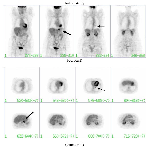
| Description | A 71-year-old lady was incidentally found to have a coin lesion on a chest X-ray. Thoracic CT confirmed the presence of a 1.0 cm solitary pulmonary nodule. The initial FDG-PET study shown here (Philips Allegro system) reveals a small retrocardiac focus of increased FDG uptake within the left lower lobe nodule (thin arrow) suggestive of a malignant process. In addition, the study demonstrates abnormal increased activity at the gastro-oesophageal junction (thick arrow). The patient was found to have non-Hodgkin’s lymphoma at both sites. |
| Date | |
| Source | Gastric and pulmonary lymphoma presenting as a solitary pulmonary nodule, Biomed Imaging Interv J 2007; 3(4):e51. doi:10.2349/biij.3.4.e51. |
| Author | Thomas EL, Lenzo NP, Troedson R |
Licensing[edit]
This file is licensed under the Creative Commons Attribution-Share Alike 3.0 Unported license.
- You are free:
- to share – to copy, distribute and transmit the work
- to remix – to adapt the work
- Under the following conditions:
- attribution – You must give appropriate credit, provide a link to the license, and indicate if changes were made. You may do so in any reasonable manner, but not in any way that suggests the licensor endorses you or your use.
- share alike – If you remix, transform, or build upon the material, you must distribute your contributions under the same or compatible license as the original.
File history
Click on a date/time to view the file as it appeared at that time.
| Date/Time | Thumbnail | Dimensions | User | Comment | |
|---|---|---|---|---|---|
| current | 20:26, 3 July 2008 | | 585 × 578 (42 KB) | Stevenfruitsmaak (talk | contribs) | {{Information |Description=A 71-year-old lady was incidentally found to have a coin lesion on a chest X-ray. Thoracic CT confirmed the presence of a 1.0 cm solitary pulmonary nodule. The initial FDG-PET study shown here (Philips Allegro system) reveals a |
You cannot overwrite this file.
File usage on Commons
The following page uses this file:
File usage on other wikis
The following other wikis use this file:
- Usage on de.wikipedia.org
- Usage on en.wikipedia.org
- Usage on it.wikipedia.org
- Usage on outreach.wikimedia.org
- Usage on ru.wikipedia.org