File:Lodi - Ospedale Vecchio 1098.jpg
Jump to navigation
Jump to search
Size of this preview: 400 × 600 pixels. Other resolutions: 160 × 240 pixels | 320 × 480 pixels | 512 × 768 pixels | 682 × 1,024 pixels | 1,365 × 2,048 pixels | 3,896 × 5,844 pixels.
Original file (3,896 × 5,844 pixels, file size: 15.38 MB, MIME type: image/jpeg)
Captions
Captions
Add a one-line explanation of what this file represents
Summary[edit]
| Description |
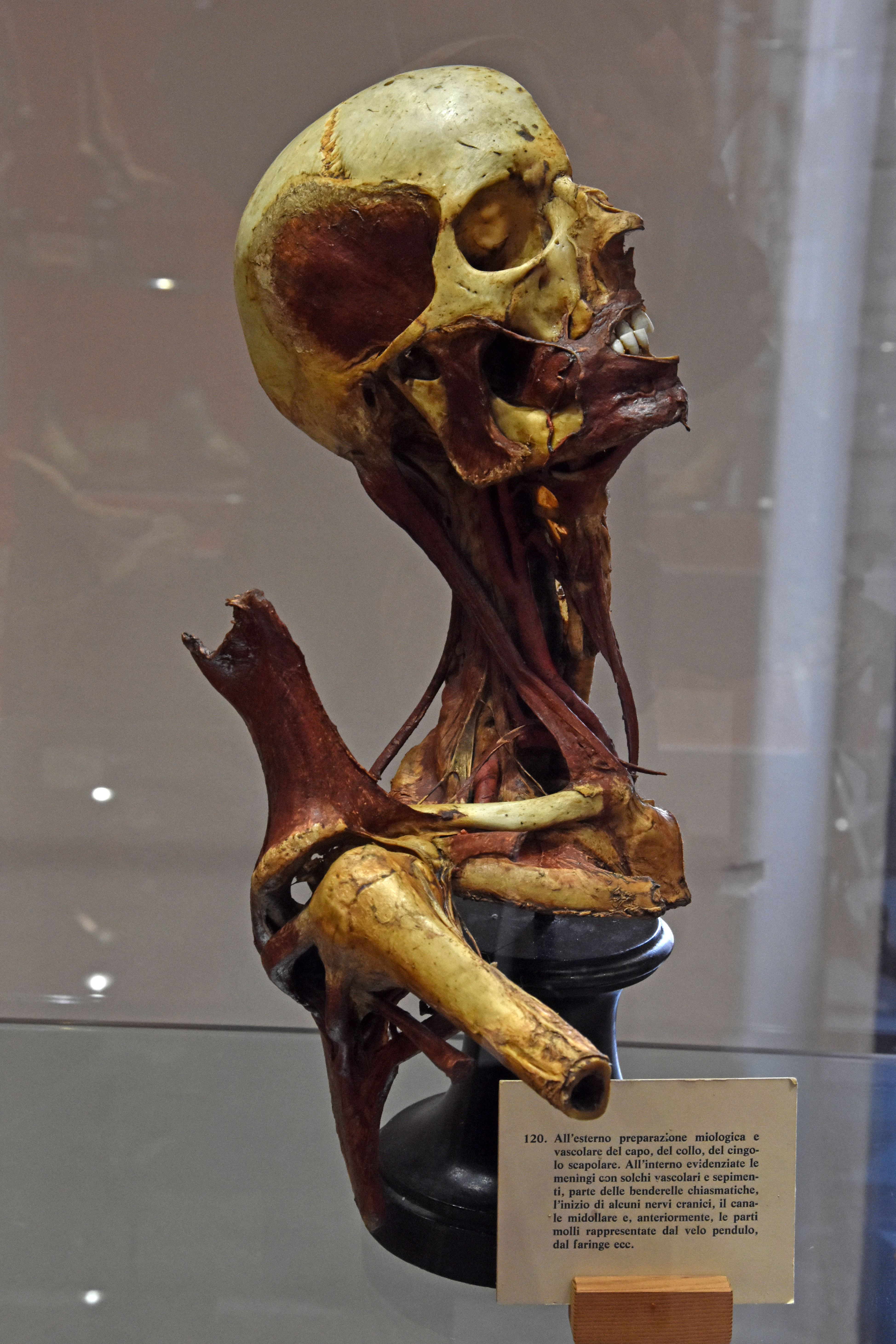
Italiano: La collezione anatomica "Paolo Gorini" nell'Ospedale Vecchio di Lodi. 120. All'esterno preparazione miologica e vascolare del capo, del collo, del cingolo scapolare. All'interno evidenziate le meningi con solchi vascolari e sepimenti, parte delle benderelle chiasmatiche, l'inizio di alcuni nervi cranici, il canale midollare e, anteriormente, le parti molli, rappresentate dal velo pendulo, dal faringe ecc. |
| Date | Taken on 12 October 2022 |
| Source | Own work |
| Author | Phyrexian |
| Camera | This photograph was taken with a Nikon D3400 |
Licensing[edit]
I, the copyright holder of this work, hereby publish it under the following license:
This file is licensed under the Creative Commons Attribution-Share Alike 4.0 International license.
- You are free:
- to share – to copy, distribute and transmit the work
- to remix – to adapt the work
- Under the following conditions:
- attribution – You must give appropriate credit, provide a link to the license, and indicate if changes were made. You may do so in any reasonable manner, but not in any way that suggests the licensor endorses you or your use.
- share alike – If you remix, transform, or build upon the material, you must distribute your contributions under the same or compatible license as the original.
File history
Click on a date/time to view the file as it appeared at that time.
| Date/Time | Thumbnail | Dimensions | User | Comment | |
|---|---|---|---|---|---|
| current | 12:22, 16 November 2022 | | 3,896 × 5,844 (15.38 MB) | Phyrexian (talk | contribs) | {{Information |description = {{it|La collezione anatomica "Paolo Gorini" nell'Ospedale Vecchio di Lodi. 120. All'esterno preparazione miologica e vascolare del capo, del collo, del cingolo scapolare. All'interno evidenziate le meningi con solchi vascolari e sepimenti, parte delle benderelle chiasmatiche, l'inizio di alcuni nervi cranici, il canale midollare e, anteriormente, le parti molli, rappresentate dal velo pendulo, dal faringe ec... |
You cannot overwrite this file.
File usage on Commons
There are no pages that use this file.