File:Schematic of Cell State Splitter Organelle.jpg
Jump to navigation
Jump to search
No higher resolution available.
Schematic_of_Cell_State_Splitter_Organelle.jpg (324 × 215 pixels, file size: 14 KB, MIME type: image/jpeg)
Captions
Captions
Add a one-line explanation of what this file represents
Summary[edit]
| Description |
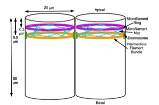
English: The cell state splitter is an organelle predicted and then first observed at the apical end of ectoderm cells in axolotl embryos at early gastrulation. It consists of an upper microfilament ring with an intermediate filament ring below it, subtended by a mat of microtubules. (Note not to scale: the vertical and horizontal scales are compressed, as 50 μm = 100 x 0.5 μm. The cell state splitter occupies only the top 1% of the height of an ectoderm cell.) |
| Date | |
| Source | Own work |
| Author | Bjorklund21 |
This work was published in my book, Embryogenesis Explained, World Scientific, Singapore, 2016, figure 6.3, page 445 and I have approval from the publisher to release it here.
Licensing[edit]
I, the copyright holder of this work, hereby publish it under the following license:
This file is licensed under the Creative Commons Attribution 4.0 International license.
- You are free:
- to share – to copy, distribute and transmit the work
- to remix – to adapt the work
- Under the following conditions:
- attribution – You must give appropriate credit, provide a link to the license, and indicate if changes were made. You may do so in any reasonable manner, but not in any way that suggests the licensor endorses you or your use.
File history
Click on a date/time to view the file as it appeared at that time.
| Date/Time | Thumbnail | Dimensions | User | Comment | |
|---|---|---|---|---|---|
| current | 19:30, 6 November 2017 | | 324 × 215 (14 KB) | Bjorklund21 (talk | contribs) | User created page with UploadWizard |
You cannot overwrite this file.
File usage on Commons
There are no pages that use this file.
File usage on other wikis
The following other wikis use this file:
- Usage on en.wikipedia.org