File:Tetraspora gelatinosa.jpg

Original file (1,047 × 700 pixels, file size: 139 KB, MIME type: image/jpeg)
Captions
Captions
Summary[edit]
| Description |
Identifier: algvolimyxophy00west Title: Algæ. Vol. I. Myxophyceæ, Peridinieæ, Bacillarieæ, Chlorophyceæ, together with a brief summary of the occurrence and distribution of freshwat4er Algæ Year: 1916 (1910s) Authors: West, G. S. (George Stephen), 1876-1919 Subjects: Algae Publisher: Cambridge [Eng.] The University press Contributing Library: MBLWHOI Library Digitizing Sponsor: MBLWHOI Library
Click here to view book online to see this illustration in context in a browseable online version of this book.
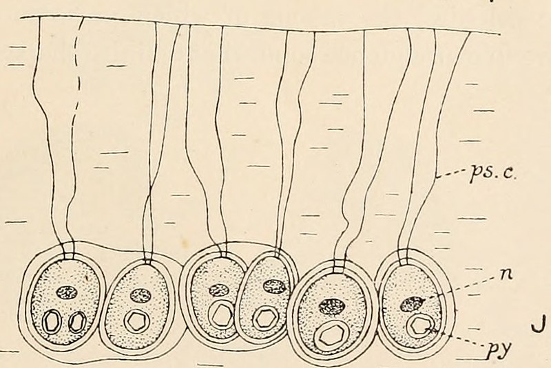
Text Appearing After Image: Fig. 113. A-G, Schizochlamys gelatinosa A. Br. A, vegetative cell showing pseudocilia, ×625; B, cell showing ecdysis of outer layers of wall, ×415; C and D, formation of zoogonidia, ×625; E, zoogonidium, ×830; F and G, zoogonidium changing to Schizochlamys-cell, ×830. H and I, Apiocystis brauniana Näg. H, pear-shaped colony, ×430; I, three cells showing pseudocilia, b, two daughter-cells from a division, the second pseudocilium not yet developed, ×860. J, Tetraspora gelatinosa (Vauch.) Desv., periphery of colony showing a few of the cells with their pseudocilia, × about 900. cv, contractile vacuole; n, nucleus; ol, oil globule; ps.c., pseudocilia; py, pyrenoid ; sf, stigma (or pigment-spot). (A-G, after Scherffel; J, after Chodat.) colony. The cells multiply by repeated division, chiefly in two directions inone plane, with the conversion of the walls of the mother-cells into mucilage.The pseudocilia are embedded in the mucilage of the colony (fig. 113 J), andeach cell is o
|
| Date | |
| Source | Image from page 200 of "Algæ. Vol. I. Myxophyceæ, Peridinieæ, Bacillarieæ, Chlorophyceæ, together with a brief summary of the occurrence and distribution of freshwat4er Algæ" (1916) |
| Author | Internet Archive Book Images |
| Permission (Reusing this file) |
Internet Archive Book Images @ Flickr Commons |
| Other versions |
.jpg)
Licensing[edit]
This image was taken from Flickr's The Commons. The uploading organization may have various reasons for determining that no known copyright restrictions exist, such as:
More information can be found at https://flickr.com/commons/usage/. Please add additional copyright tags to this image if more specific information about copyright status can be determined. See Commons:Licensing for more information. |
Licensing[edit]
|
This work is in the public domain in its country of origin and other countries and areas where the copyright term is the author's life plus 100 years or fewer. This work is in the public domain in the United States because it was published (or registered with the U.S. Copyright Office) before January 1, 1929. | |
| This file has been identified as being free of known restrictions under copyright law, including all related and neighboring rights. | |
File history
Click on a date/time to view the file as it appeared at that time.
| Date/Time | Thumbnail | Dimensions | User | Comment | |
|---|---|---|---|---|---|
| current | 14:06, 3 October 2019 | | 1,047 × 700 (139 KB) | Awkwafaba (talk | contribs) | File:Image from page 200 of "Algæ. Vol. I. Myxophyceæ, Peridinieæ, Bacillarieæ, Chlorophyceæ, together with a brief summary of the occurrence and distribution of freshwat4er Algæ" (1916).jpg cropped 35 % horizontally, 57 % vertically using CropTool with lossless mode. |
You cannot overwrite this file.
File usage on Commons
The following 2 pages use this file:
File usage on other wikis
The following other wikis use this file:
- Usage on ceb.wikipedia.org
- Usage on fr.wikipedia.org
- Usage on species.wikimedia.org
- Usage on tr.wikipedia.org
- Usage on www.wikidata.org
- Usage on zh.wikipedia.org