File:Villous adenoma of the sigmoid colon 1.jpg
From Wikimedia Commons, the free media repository
Jump to navigation
Jump to search
Villous_adenoma_of_the_sigmoid_colon_1.jpg (550 × 350 pixels, file size: 54 KB, MIME type: image/jpeg)
File information
Structured data
Captions
Captions
Add a one-line explanation of what this file represents
Summary[edit]
| DescriptionVillous adenoma of the sigmoid colon 1.jpg |
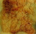
Villous adenoma This large (6.5 cm) villous adenoma of the sigmoid colon had no invasive component, so the patient's prognosis after excision is excellent. The photo above is shot using conventional copy stand techniques and shows what this tumor looked like to the pathologist on the gross board, and to the gastroenterologist through the endoscope. The photos below show the specimen as immersed in tapwater. The buoyancy of the delicate villous structures causes them to stand up and separate, showing the complex coral-like filigree produced by this type of growth pattern. |
| Date | Posted 27 May 01; updated 20 Feb 05 |
| Source | http://web2.airmail.net/uthman/specimens/index.html |
| Author | Photography by Ed Uthman, MD. |
| Permission (Reusing this file) |
Public domain. |
| Other versions |
|
Licensing[edit]
| Public domainPublic domainfalsefalse |
| This work has been released into the public domain by its author, Ed Uthman. This applies worldwide. In some countries this may not be legally possible; if so: Ed Uthman grants anyone the right to use this work for any purpose, without any conditions, unless such conditions are required by law. Public domainPublic domainfalsefalse |
File history
Click on a date/time to view the file as it appeared at that time.
| Date/Time | Thumbnail | Dimensions | User | Comment | |
|---|---|---|---|---|---|
| current | 23:26, 4 June 2006 |  | 550 × 350 (54 KB) | Patho (talk | contribs) | {{Information| |Description=Villous adenoma villous adenoma This large (6.5 cm) villous adenoma of the sigmoid colon had no invasive component, so the patient's prognosis after excision is excellent. The photo above is shot using conventional copy stand |
You cannot overwrite this file.
File usage on Commons
The following 3 pages use this file:
File usage on other wikis
The following other wikis use this file:
- Usage on de.wikibooks.org
Metadata
This file contains additional information such as Exif metadata which may have been added by the digital camera, scanner, or software program used to create or digitize it. If the file has been modified from its original state, some details such as the timestamp may not fully reflect those of the original file. The timestamp is only as accurate as the clock in the camera, and it may be completely wrong.
| _error | 0 |
|---|


