Category:Anatomy of the human eye
Saltar para a navegação
Saltar para a pesquisa
categoria de um projeto da Wikimedia | |||||
| Carregar ficheiro | |||||
| Instância de | |||||
|---|---|---|---|---|---|
| |||||
Subcategorias
Esta categoria contém as seguintes 15 subcategorias (de um total de 15).
C
- Human conjunctiva (2 F)
D
E
- Extraocular muscles (42 F)
- Anatomy of the human eyelids (12 F)
H
L
M
- Meibomian gland (4 F)
N
O
P
- Plica semilunaris (9 F)
S
- Human surface anatomy of eye (14 F)
V
Páginas na categoria "Anatomy of the human eye"
Esta categoria só contém a seguinte página.
Multimédia na categoria "Anatomy of the human eye"
Esta categoria contém os seguintes 154 ficheiros (de um total de 154).
-
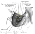
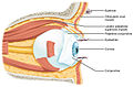 1411 Eye in The Orbit.jpg 1 742 × 1 140; 808 kB
-
 A Series of Anatomical Plates Nerves Plate 29.jpg 800 × 1 055; 164 kB
-
 A szivárványhártya szerkezete.png 1 186 × 1 175; 706 kB
-
-
 Akis.jpg 800 × 600; 49 kB
-

-
 Anatomie oka 01.jpg 9 248 × 6 944; 14,89 MB
-
 Anatomie oka 02.jpg 9 248 × 6 944; 15,26 MB
-
 Anatomie oka 03.jpg 9 248 × 6 944; 15,43 MB
-
 Anatomy of the eye, in Iconum Anatomicarum. Wellcome M0017124.jpg 3 898 × 2 864; 2,5 MB
-
 Anatomy of the eye. Wellcome M0001658.jpg 1 494 × 1 431; 574 kB
-
 Arizona eye model.png 921 × 410; 38 kB
-
 Augen mit Sehnerven von oben (Meyers).jpg 605 × 491; 107 kB
-
 Augenbewegung.jpg 200 × 139; 10 kB
-
 Augennerven.jpg 663 × 420; 96 kB
-
 Zur Anatomie der gesunden und kranken Linse - von Otto Becker ; unter Mitwirkung von J. R. Da Gama Pinto und H. Schäfer. (IA b21641870).pdf 1 379 × 2 056, 158 páginas; 17,17 MB
-
 Ball Collection, Acc 21818 (3175211588).jpg 1 800 × 2 501; 2,68 MB
-
 Ball Collection, Acc 21834.jpg 497 × 800; 57 kB
-
 Begiaren anatomiaren modeloa.png 3 274 × 2 855; 6,91 MB
-
 Brockhaus and Efron Encyclopedic Dictionary b16 812-0.jpg 1 582 × 2 577; 462 kB
-
 Choroid.jpg 243 × 207; 15 kB
-
 Co-2-435f2.jpg 465 × 278; 67 kB
-
 Color perception.jpg 446 × 270; 32 kB
-
 DBP 1994 1752 Hermann von Helmholtz.jpg 1 070 × 670; 391 kB
-
 Demonstration model of the human eye, French. 1884 Wellcome S0008222.jpg 1 525 × 1 268; 749 kB
-
 Density rods n cones.png 598 × 585; 117 kB
-
 Descartes body physics 1.jpg 887 × 606; 221 kB
-
 Descartes body physics 2.jpg 947 × 611; 246 kB
-

-
 Detail of gnomonic projection.png 856 × 331; 38 kB
-

-

-
 Distribution of Cones and Rods on Human Retina sCH.png 891 × 557; 13 kB
-
 Ear and eye; eight figures, including cross-section of eye. Wellcome V0007959.jpg 3 114 × 3 936; 2,31 MB
-
 EB1911 Vision - Ideal or Schematique Eye.jpg 937 × 585; 177 kB
-
 EB1911 Vision - Mechanism of Accommodation.jpg 963 × 513; 119 kB
-

-
 Extraocular muscles.jpg 960 × 720; 99 kB
-
 Eye (11291008035).jpg 2 211 × 1 512; 423 kB
-
 Eye Central Heterochromia (2).jpg 1 122 × 614; 439 kB
-
 Eye Full Work.jpg 2 776 × 1 404; 1,05 MB
-
 Eye illustration, 17th century Wellcome M0011407.jpg 2 480 × 4 280; 3,28 MB
-
 Eye Line of sight.jpg 287 × 208; 20 kB
-
 Eye lines of sight.png 833 × 602; 785 kB
-
 Eye orbit anatomy anterior.jpg 2 934 × 1 924; 1 MB
-
 Eye orbit anatomy anterior2.jpg 2 934 × 1 924; 3,71 MB
-
 Eye orbit anterior (modified).jpg 1 400 × 933; 301 kB
-
 Eye orbit anterior.jpg 1 400 × 933; 1,33 MB
-
 Eye Vision fields.jpg 1 664 × 2 060; 730 kB
-
 Eye, 17th century Wellcome L0007983.jpg 2 598 × 3 828; 3,66 MB
-
 Eye, 17th century Wellcome L0007985.jpg 1 036 × 1 696; 778 kB
-
 Eye-diagram no circles border.svg 1 237 × 1 208; 130 kB
-
 EyeMuscles.gif 600 × 389; 48 kB
-
 Eyes from "Anatomia Humani Corporis", Bidloo, 1685 Wellcome L0013456.jpg 1 110 × 1 704; 669 kB
-
 Eyesheaths.jpg 550 × 499; 72 kB
-
 Fig 4 PSWG 1920.gif 544 × 501; 24 kB
-

-
 Fotothek df tg 0001900 Optik ^ Anatomie ^ Mensch ^ Auge.jpg 800 × 544; 235 kB
-
 Fotothek df tg 0001903 Optik ^ Anatomie ^ Auge ^ Mensch.jpg 800 × 525; 195 kB
-
 Fotothek df tg 0001919 Optik ^ Anatomie ^ Mensch ^ Auge.jpg 692 × 820; 334 kB
-
 Fotothek df tg 0003714 Optik ^ Biologie ^ Auge ^ Mensch.jpg 535 × 820; 154 kB
-
 Fotothek df tg 0003715 Optik ^ Lochkamera.jpg 800 × 598; 158 kB
-

-
 Fotothek df tg 0006586 Biologie ^ Anatomie ^ Mensch.jpg 489 × 820; 184 kB
-
 Fotothek df tg 0006588 Biologie ^ Anatomie ^ Mensch.jpg 378 × 820; 136 kB
-
 GDX - Abweichungsdarstellung.png 692 × 645; 401 kB
-
 GDx - Fundusbild.png 692 × 645; 640 kB
-
 Gray164- Fosse du sac lacrymal.jpg 400 × 379; 114 kB
-
 Gray776.png 353 × 650; 61 kB
-
 Gray777.png 700 × 455; 60 kB
-
 Gray785.png 348 × 500; 33 kB
-
 Gray787.png 413 × 400; 36 kB
-
 Gray888 zh.png 550 × 464; 197 kB
-
 Gray888.png 550 × 464; 67 kB
-
 Göttingen-Augenmodell.JPG 2 136 × 2 848; 1,87 MB
-
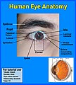 Human eye anatomy.jpg 4 320 × 4 800; 3,77 MB
-
 Human Eye Model (50692872268).jpg 2 756 × 2 417; 942 kB
-
 Human Eye Model (50692874163).jpg 4 086 × 2 702; 2,09 MB
-
 Human Eye Model (50693611856).jpg 3 210 × 2 757; 1,67 MB
-
 Human Eye Model (50693612761).jpg 3 326 × 2 512; 1,44 MB
-
 Human Eye Model (50693614536).jpg 3 563 × 2 716; 1,59 MB
-
 Human Eye Model (50693720447).jpg 3 576 × 3 014; 1,97 MB
-
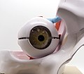
 Human eyeball.jpg 2 368 × 3 537; 3,36 MB
-
 Iconographic Encyclopedia of Science, Literature and Art 153.jpg 2 851 × 2 278; 1,28 MB
-
 Iris structure.png 1 259 × 1 355; 907 kB
-
 Ivory and horn model of an eye, Europe, 1601-1700 Wellcome L0058730.jpg 2 748 × 3 964; 742 kB
-
 Ivory and horn model of an eye, Europe, 1801-1900 Wellcome L0057567.jpg 4 256 × 2 832; 810 kB
-
 LA2-NSRW-2-0169.jpg 1 866 × 2 797; 1,15 MB
-
-
 Lateral orbit anatomy 2.jpg 1 915 × 1 250; 1,13 MB
-
 Lateral orbit nerves.jpg 1 915 × 1 250; 917 kB
-
 Lehrbuch der Augenheilkunde (1891) (14760827241).jpg 1 539 × 3 240; 807 kB
-
 Meyers b2 s0074a.jpg 2 048 × 1 608; 609 kB
-
 Meyers b2 s0078a.jpg 2 048 × 1 657; 295 kB
-
 Meyers b7 s0235.jpg 800 × 1 275; 684 kB
-

-
 Model of an eye, Europe, 1801-1900 Wellcome L0058736.jpg 2 832 × 4 012; 1,18 MB
-
 Model of the eye in 4 parts by William Rush.jpg 3 982 × 3 188; 1,48 MB
-
 Modell vom menschlichen Auge.jpg 1 000 × 1 592; 292 kB
-

-
 NEI laboratory research eye.jpg 4 034 × 2 691; 4,46 MB
-
 Nerv.jpg 770 × 396; 318 kB
-
 Optic nerve and ocular muscles. Wellcome L0009855.jpg 1 604 × 1 222; 861 kB
-
 Orbital septum.png 917 × 832; 245 kB
-
 Orléans - MOBE 22.jpg 3 648 × 5 472; 4,63 MB
-
 Page from An atlas of anatomical plates Wellcome L0067250.jpg 4 795 × 7 671; 6,22 MB
-
 Planetarium (4).jpg 1 024 × 768; 210 kB
-
 Plaster model of a section of the human eye Wellcome L0035459.jpg 1 908 × 2 412; 784 kB
-
 Plaster model of a section of the human eye Wellcome L0035460.jpg 1 900 × 2 384; 666 kB
-
 Plaster model of a section of the human eye Wellcome L0035462.jpg 1 916 × 2 876; 948 kB
-
 Prisma eye sensors 02.jpg 50 × 188; 8 kB
-
 Prisma eye sensors 03.jpg 50 × 234; 9 kB
-
 Prisma eye sensors.jpg 50 × 216; 9 kB
-
 PSM V45 D214 Human eyeball with outer wall of orbit removed.jpg 1 581 × 1 083; 291 kB
-
 PSM V45 D228 Image at the focus of a lens.jpg 1 739 × 1 730; 214 kB
-
 PSM V45 D230 Image at the focus of a concave mirror.jpg 1 681 × 1 685; 151 kB
-
 PSM V45 D232 Achromatic object glass.jpg 818 × 751; 30 kB
-
 Purkinje Tree.png 282 × 123; 18 kB
-
 Receptive field sCH.png 403 × 737; 52 kB
-
 Receptive field-ar.png 403 × 737; 57 kB
-
 Receptive field.png 403 × 737; 67 kB
-
 Representacion pictorica del codigo del iris.png 923 × 189; 86 kB
-
 Result-of-the-sample-pupil-region-detection.jpg 600 × 460; 128 kB
-
 Retinal pigment epithelium.jpg 1 602 × 1 799; 1,2 MB
-
 Rez lid rohovkou.gif 800 × 581; 138 kB
-
 Rez rohovkou.png 3 509 × 2 550; 9,63 MB
-
 Rohovka vrstvy.gif 350 × 229; 25 kB
-
 Schematic diagram of the human eye be.svg 449 × 423; 264 kB
-
 Schematic diagram of the human eye ru.svg 449 × 423; 265 kB
-
 Sobo 1909 752.png 1 326 × 1 038; 3,95 MB
-
 Sobo 1909 758.png 1 590 × 1 155; 5,26 MB
-
 Sobo 1909 759.png 1 437 × 954; 3,93 MB
-
 Sobo 1909 761.png 1 485 × 1 047; 4,46 MB
-
 Sobo 1909 762.png 1 773 × 1 212; 6,16 MB
-
 Sobo 1909 763.png 1 752 × 1 140; 5,72 MB
-
 Sobo 1911 746.png 2 368 × 1 484; 10,07 MB
-
 Sobo 1911 747.png 1 964 × 1 080; 6,08 MB
-
 Sobo 1911 750.png 2 188 × 1 292; 8,1 MB
-

-
 Spirale de Tilllaux.jpg 1 134 × 868; 278 kB
-
 The anatomy of the eyes and optic nerve. Wellcome M0011340.jpg 3 791 × 2 787; 2,62 MB
-
-

-

-

-
 Trochlear and frontal nerves.jpg 960 × 720; 100 kB
-
 Visible-human-eye.jpg 2 188 × 1 230; 347 kB
-
Wie ist ein Auge aufgebaut (CC BY 4.0) .webm 32 s, 1 280 × 720; 2,82 MB
-
 Woodcuts; anatomy of the eye, circa 1503. Wellcome M0010695.jpg 2 880 × 3 726; 2,02 MB
-
 Σχηματικό διάγραμμα ανθρώπινου ματιού.png 1 123 × 1 294; 287 kB
-

-
 Инфографика-анатомия органа зрения.png 1 840 × 2 376; 415 kB
-
 Колбочки.jpg 471 × 421; 52 kB
-
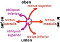
 Мускули на окото.jpg 974 × 302; 77 kB


_(14802978143).jpg)


.jpg)


.jpg)


_021_Die_kn%C3%B6cherne_Augenh%C3%B6hle_mit_dem_Augapfel_(rechts).png)
_022_Senkrechter_Schnitt_durch_die_Augenh%C3%B6hle.png)


_(14583575838).jpg)

.jpg)
.jpg)


.jpg)


.jpg)
.jpg)
.jpg)
.jpg)
.jpg)
.jpg)


_(14760827241).jpg)


_(14580250677).jpg)


.jpg)


_(14597798710).jpg)


_(14779540115).jpg)


