Category:Anatomy of the human eye
Jump to navigation
Jump to search
Wikimedia category | |||||
| Upload media | |||||
| Instance of | |||||
|---|---|---|---|---|---|
| |||||
Subcategories
This category has the following 15 subcategories, out of 15 total.
C
- Human conjunctiva (2 F)
D
E
- Extraocular muscles (43 F)
- Anatomy of the human eyelids (12 F)
H
L
M
- Meibomian gland (4 F)
N
O
P
- Plica semilunaris (9 F)
S
- Human surface anatomy of eye (14 F)
V
Pages in category "Anatomy of the human eye"
This category contains only the following page.
Media in category "Anatomy of the human eye"
The following 155 files are in this category, out of 155 total.
-
 1411 Eye in The Orbit.jpg 1,742 × 1,140; 808 KB
-
 A Series of Anatomical Plates Nerves Plate 29.jpg 800 × 1,055; 164 KB
-
 A szivárványhártya szerkezete.png 1,186 × 1,175; 706 KB
-
-
 Akis.jpg 800 × 600; 49 KB
-

-
 Anatomie oka 01.jpg 9,248 × 6,944; 14.89 MB
-
 Anatomie oka 02.jpg 9,248 × 6,944; 15.26 MB
-
 Anatomie oka 03.jpg 9,248 × 6,944; 15.43 MB
-
 Anatomy of the eye, in Iconum Anatomicarum. Wellcome M0017124.jpg 3,898 × 2,864; 2.5 MB
-
 Anatomy of the eye. Wellcome M0001658.jpg 1,494 × 1,431; 574 KB
-
 Arizona eye model.png 921 × 410; 38 KB
-
 Augen mit Sehnerven von oben (Meyers).jpg 605 × 491; 107 KB
-
 Augenbewegung.jpg 200 × 139; 10 KB
-
 Augennerven.jpg 663 × 420; 96 KB
-
 Zur Anatomie der gesunden und kranken Linse - von Otto Becker ; unter Mitwirkung von J. R. Da Gama Pinto und H. Schäfer. (IA b21641870).pdf 1,379 × 2,056, 158 pages; 17.17 MB
-
 Ball Collection, Acc 21818 (3175211588).jpg 1,800 × 2,501; 2.68 MB
-
 Ball Collection, Acc 21834.jpg 497 × 800; 57 KB
-
 Begiaren anatomiaren modeloa.png 3,274 × 2,855; 6.91 MB
-
 Brockhaus and Efron Encyclopedic Dictionary b16 812-0.jpg 1,582 × 2,577; 462 KB
-
 Choroid.jpg 243 × 207; 15 KB
-
 Co-2-435f2.jpg 465 × 278; 67 KB
-
 Color perception.jpg 446 × 270; 32 KB
-
 DBP 1994 1752 Hermann von Helmholtz.jpg 1,070 × 670; 391 KB
-
 Demonstration model of the human eye, French. 1884 Wellcome S0008222.jpg 1,525 × 1,268; 749 KB
-
 Density rods n cones.png 598 × 585; 117 KB
-
 Descartes body physics 1.jpg 887 × 606; 221 KB
-
 Descartes body physics 2.jpg 947 × 611; 246 KB
-

-
 Detail of gnomonic projection.png 856 × 331; 38 KB
-

-

-
 Distribution of Cones and Rods on Human Retina sCH.png 891 × 557; 13 KB
-
 Ear and eye; eight figures, including cross-section of eye. Wellcome V0007959.jpg 3,114 × 3,936; 2.31 MB
-
 EB1911 Vision - Ideal or Schematique Eye.jpg 937 × 585; 177 KB
-
 EB1911 Vision - Mechanism of Accommodation.jpg 963 × 513; 119 KB
-

-
 Extraocular Eye Muscles.png 600 × 389; 194 KB
-
 Extraocular muscles.jpg 960 × 720; 99 KB
-
 Eye (11291008035).jpg 2,211 × 1,512; 423 KB
-
 Eye Central Heterochromia (2).jpg 1,122 × 614; 439 KB
-
 Eye Full Work.jpg 2,776 × 1,404; 1.05 MB
-
 Eye illustration, 17th century Wellcome M0011407.jpg 2,480 × 4,280; 3.28 MB
-
 Eye Line of sight.jpg 287 × 208; 20 KB
-
 Eye lines of sight.png 833 × 602; 785 KB
-
 Eye orbit anatomy anterior.jpg 2,934 × 1,924; 1 MB
-
 Eye orbit anatomy anterior2.jpg 2,934 × 1,924; 3.71 MB
-
 Eye orbit anterior (modified).jpg 1,400 × 933; 301 KB
-
 Eye orbit anterior.jpg 1,400 × 933; 1.33 MB
-
 Eye Vision fields.jpg 1,664 × 2,060; 730 KB
-
 Eye, 17th century Wellcome L0007983.jpg 2,598 × 3,828; 3.66 MB
-
 Eye, 17th century Wellcome L0007985.jpg 1,036 × 1,696; 778 KB
-
 Eye-diagram no circles border.svg 1,237 × 1,208; 130 KB
-
 EyeMuscles.gif 600 × 389; 48 KB
-
 Eyes from "Anatomia Humani Corporis", Bidloo, 1685 Wellcome L0013456.jpg 1,110 × 1,704; 669 KB
-
 Eyesheaths.jpg 550 × 499; 72 KB
-
 Fig 4 PSWG 1920.gif 544 × 501; 24 KB
-

-
 Fotothek df tg 0001900 Optik ^ Anatomie ^ Mensch ^ Auge.jpg 800 × 544; 235 KB
-
 Fotothek df tg 0001903 Optik ^ Anatomie ^ Auge ^ Mensch.jpg 800 × 525; 195 KB
-
 Fotothek df tg 0001919 Optik ^ Anatomie ^ Mensch ^ Auge.jpg 692 × 820; 334 KB
-
 Fotothek df tg 0003714 Optik ^ Biologie ^ Auge ^ Mensch.jpg 535 × 820; 154 KB
-
 Fotothek df tg 0003715 Optik ^ Lochkamera.jpg 800 × 598; 158 KB
-

-
 Fotothek df tg 0006586 Biologie ^ Anatomie ^ Mensch.jpg 489 × 820; 184 KB
-
 Fotothek df tg 0006588 Biologie ^ Anatomie ^ Mensch.jpg 378 × 820; 136 KB
-
 GDX - Abweichungsdarstellung.png 692 × 645; 401 KB
-
 GDx - Fundusbild.png 692 × 645; 640 KB
-
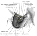
 Gray164- Fosse du sac lacrymal.jpg 400 × 379; 114 KB
-
 Gray776.png 353 × 650; 61 KB
-
 Gray777.png 700 × 455; 60 KB
-
 Gray785.png 348 × 500; 33 KB
-
 Gray787.png 413 × 400; 36 KB
-
 Gray888 zh.png 550 × 464; 197 KB
-
 Gray888.png 550 × 464; 67 KB
-
 Göttingen-Augenmodell.JPG 2,136 × 2,848; 1.87 MB
-
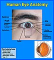 Human eye anatomy.jpg 4,320 × 4,800; 3.77 MB
-
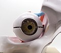 Human Eye Model (50692872268).jpg 2,756 × 2,417; 942 KB
-
 Human Eye Model (50692874163).jpg 4,086 × 2,702; 2.09 MB
-
 Human Eye Model (50693611856).jpg 3,210 × 2,757; 1.67 MB
-
 Human Eye Model (50693612761).jpg 3,326 × 2,512; 1.44 MB
-
 Human Eye Model (50693614536).jpg 3,563 × 2,716; 1.59 MB
-
 Human Eye Model (50693720447).jpg 3,576 × 3,014; 1.97 MB
-
 Human eyeball.jpg 2,368 × 3,537; 3.36 MB
-
 Iconographic Encyclopedia of Science, Literature and Art 153.jpg 2,851 × 2,278; 1.28 MB
-
 Iris structure.png 1,259 × 1,355; 907 KB
-
 Ivory and horn model of an eye, Europe, 1601-1700 Wellcome L0058730.jpg 2,748 × 3,964; 742 KB
-
 Ivory and horn model of an eye, Europe, 1801-1900 Wellcome L0057567.jpg 4,256 × 2,832; 810 KB
-
 LA2-NSRW-2-0169.jpg 1,866 × 2,797; 1.15 MB
-
-
 Lateral orbit anatomy 2.jpg 1,915 × 1,250; 1.13 MB
-
 Lateral orbit nerves.jpg 1,915 × 1,250; 917 KB
-
 Lehrbuch der Augenheilkunde (1891) (14760827241).jpg 1,539 × 3,240; 807 KB
-
 Meyers b2 s0074a.jpg 2,048 × 1,608; 609 KB
-
 Meyers b2 s0078a.jpg 2,048 × 1,657; 295 KB
-
 Meyers b7 s0235.jpg 800 × 1,275; 684 KB
-

-
 Model of an eye, Europe, 1801-1900 Wellcome L0058736.jpg 2,832 × 4,012; 1.18 MB
-
 Model of the eye in 4 parts by William Rush.jpg 3,982 × 3,188; 1.48 MB
-
 Modell vom menschlichen Auge.jpg 1,000 × 1,592; 292 KB
-

-
 NEI laboratory research eye.jpg 4,034 × 2,691; 4.46 MB
-
 Nerv.jpg 770 × 396; 318 KB
-
 Optic nerve and ocular muscles. Wellcome L0009855.jpg 1,604 × 1,222; 861 KB
-
 Orbital septum.png 917 × 832; 245 KB
-
 Orléans - MOBE 22.jpg 3,648 × 5,472; 4.63 MB
-
 Page from An atlas of anatomical plates Wellcome L0067250.jpg 4,795 × 7,671; 6.22 MB
-
 Planetarium (4).jpg 1,024 × 768; 210 KB
-
 Plaster model of a section of the human eye Wellcome L0035459.jpg 1,908 × 2,412; 784 KB
-
 Plaster model of a section of the human eye Wellcome L0035460.jpg 1,900 × 2,384; 666 KB
-
 Plaster model of a section of the human eye Wellcome L0035462.jpg 1,916 × 2,876; 948 KB
-
 Prisma eye sensors 02.jpg 50 × 188; 8 KB
-
 Prisma eye sensors 03.jpg 50 × 234; 9 KB
-
 Prisma eye sensors.jpg 50 × 216; 9 KB
-
 PSM V45 D214 Human eyeball with outer wall of orbit removed.jpg 1,581 × 1,083; 291 KB
-
 PSM V45 D228 Image at the focus of a lens.jpg 1,739 × 1,730; 214 KB
-
 PSM V45 D230 Image at the focus of a concave mirror.jpg 1,681 × 1,685; 151 KB
-
 PSM V45 D232 Achromatic object glass.jpg 818 × 751; 30 KB
-
 Purkinje Tree.png 282 × 123; 18 KB
-
 Receptive field sCH.png 403 × 737; 52 KB
-
 Receptive field-ar.png 403 × 737; 57 KB
-
 Receptive field.png 403 × 737; 67 KB
-
 Representacion pictorica del codigo del iris.png 923 × 189; 86 KB
-
 Result-of-the-sample-pupil-region-detection.jpg 600 × 460; 128 KB
-
 Retinal pigment epithelium.jpg 1,602 × 1,799; 1.2 MB
-
 Rez lid rohovkou.gif 800 × 581; 138 KB
-
 Rez rohovkou.png 3,509 × 2,550; 9.63 MB
-
 Rohovka vrstvy.gif 350 × 229; 25 KB
-
 Schematic diagram of the human eye be.svg 449 × 423; 264 KB
-
 Schematic diagram of the human eye ru.svg 449 × 423; 265 KB
-
 Sobo 1909 752.png 1,326 × 1,038; 3.95 MB
-
 Sobo 1909 758.png 1,590 × 1,155; 5.26 MB
-
 Sobo 1909 759.png 1,437 × 954; 3.93 MB
-
 Sobo 1909 761.png 1,485 × 1,047; 4.46 MB
-
 Sobo 1909 762.png 1,773 × 1,212; 6.16 MB
-
 Sobo 1909 763.png 1,752 × 1,140; 5.72 MB
-
 Sobo 1911 746.png 2,368 × 1,484; 10.07 MB
-
 Sobo 1911 747.png 1,964 × 1,080; 6.08 MB
-
 Sobo 1911 750.png 2,188 × 1,292; 8.1 MB
-

-
 Spirale de Tilllaux.jpg 1,134 × 868; 278 KB
-
 The anatomy of the eyes and optic nerve. Wellcome M0011340.jpg 3,791 × 2,787; 2.62 MB
-
-

-

-

-
 Trochlear and frontal nerves.jpg 960 × 720; 100 KB
-
 Visible-human-eye.jpg 2,188 × 1,230; 347 KB
-
Wie ist ein Auge aufgebaut (CC BY 4.0) .webm 32 s, 1,280 × 720; 2.82 MB
-
 Woodcuts; anatomy of the eye, circa 1503. Wellcome M0010695.jpg 2,880 × 3,726; 2.02 MB
-
 Σχηματικό διάγραμμα ανθρώπινου ματιού.png 1,123 × 1,294; 287 KB
-

-
 Инфографика-анатомия органа зрения.png 1,840 × 2,376; 415 KB
-
 Колбочки.jpg 471 × 421; 52 KB
-
 Мускули на окото.jpg 974 × 302; 77 KB


_(14802978143).jpg)


.jpg)


.jpg)


_021_Die_kn%C3%B6cherne_Augenh%C3%B6hle_mit_dem_Augapfel_(rechts).png)
_022_Senkrechter_Schnitt_durch_die_Augenh%C3%B6hle.png)


_(14583575838).jpg)


.jpg)
.jpg)


.jpg)


.jpg)
.jpg)
.jpg)
.jpg)
.jpg)
.jpg)


_(14760827241).jpg)


_(14580250677).jpg)


.jpg)


_(14597798710).jpg)


_(14779540115).jpg)


