Category:Biochemistry methods
Saltar para a navegação
Saltar para a pesquisa
technical procedure | |||||
| Carregar ficheiro | |||||
| Subclasse de |
| ||||
|---|---|---|---|---|---|
| |||||
Subcategorias
Esta categoria contém as seguintes 19 subcategorias (de um total de 19).
B
- Benedict's reagent (10 F)
- Biuret test (12 F)
- BLOSUM (5 F)
F
H
I
- Immobilized cells (12 F)
M
- Molisch test (4 F)
O
- Open flow microperfusion (5 F)
P
R
- Reactions of ninhydrin (11 F)
T
- Tandem affinity purification (3 F)
Multimédia na categoria "Biochemistry methods"
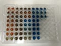
Esta categoria contém os seguintes 144 ficheiros (de um total de 144).
-
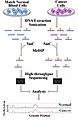
 16S PCR DGGE.jpg 499 × 273; 27 kB
-
 202202 Protein purification colored.svg 512 × 512; 33 kB
-
 202202 Protein purification monochrome.svg 512 × 512; 31 kB
-
 7-AAD.png 395 × 114; 6 kB
-
 ACGH profile of the IMR32 neuroblastoma cell line.svg 407 × 214; 4,07 MB
-
 ADN Pol E. coli.png 844 × 291; 67 kB
-
 Affinity-column-2010-09-04.png 549 × 599; 50 kB
-
 Affinity-column.png 500 × 950; 48 kB
-
 Affinity1.jpg 426 × 415; 20 kB
-
 Affinityelectrophoreses1.jpg 2 490 × 1 359; 616 kB
-
 Affinität Säule.jpg 425 × 938; 44 kB
-
 Affinitäts- Säule.png 500 × 950; 74 kB
-
 Amersham Pharmacia Biotech chromotography skid.jpg 2 322 × 4 128; 1,31 MB
-
 Amine-PEG2-Biotin.svg 512 × 170; 27 kB
-
 Amine-PEG3-Biotin.svg 512 × 149; 29 kB
-
 AptamerBinding.png 881 × 668; 186 kB
-
 Array-CGH protocol.svg 993 × 531; 2,51 MB
-
 BCIP reaction.png 4 412 × 1 015; 37 kB
-
 BCIP reaction.svg 727 × 140; 51 kB
-
 Bench-scale Bioreactor.JPG 960 × 1 280; 315 kB
-
 Biosensör bileşenleri.png 850 × 500; 655 kB
-
 Bradford assay.jpg 1 456 × 2 592; 931 kB
-
 Bradford protein assay.jpg 3 264 × 2 448; 681 kB
-
 Capillary blot setup.pdf 885 × 339; 20 kB
-
 Carlsberg micropipettes.jpg 2 383 × 1 069; 394 kB
-
 ChIP procedure.jpg 713 × 893; 51 kB
-
 Chrom SephG-50.tif 720 × 540; 78 kB
-
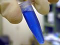 Coomassie solution.jpg 2 288 × 1 712; 2,23 MB
-
 Coomassiegel.JPG 875 × 662; 171 kB
-
 CoT analysis.png 856 × 652; 16 kB
-
 CrossedimmunoelectrophoresisofHuSe 03.jpg 309 × 250; 14 kB
-
 CrossedimmunoelectrophoresisTCBH2.jpg 1 374 × 1 569; 440 kB
-
 DamID concept.jpg 1 836 × 972; 227 kB
-
 Denovo.gif 601 × 393; 6 kB
-
 Dideoxy termination of DNA elongation EN.png 1 523 × 1 513; 34 kB
-
 Dideoxy termination of DNA elongation PL.png 1 526 × 1 514; 35 kB
-
 Didesoxy-Methode.svg 585 × 425; 107 kB
-
 DLS german.svg 552 × 439; 37 kB
-
 DLS.svg 552 × 439; 62 kB
-
 EC 1.1.1.152 reaction.PNG 2 552 × 605; 48 kB
-
 EC 1.13.11.25 reaction.PNG 1 849 × 1 164; 60 kB
-
 Edman degradation123.png 734 × 454; 8 kB
-
 Estrogen receptor assay (1).jpg 1 800 × 2 700; 2,54 MB
-
 Estrogen receptor assay (2).jpg 1 800 × 2 700; 2,46 MB
-
 Estrogen receptor assay (3).jpg 2 700 × 1 800; 1,92 MB
-
 Estrogen receptor assay (4).jpg 1 800 × 2 700; 1,6 MB
-
 Estrogen receptor assay.jpg 1 800 × 2 700; 1,98 MB
-
 Final copy.jpg 810 × 1 708; 322 kB
-
 FlexArrayer.jpg 1 918 × 1 355; 90 kB
-
 Flow-FISH 1.JPG 820 × 550; 46 kB
-
 Flow-FISH 2.JPG 930 × 699; 53 kB
-
 Flow2.jpg 800 × 3 225; 858 kB
-
 Fluorescence resonance energy transfer.jpg 709 × 547; 115 kB
-
 FRET Jablonski diagram.svg 733 × 715; 208 kB
-
 FRET-Phosgene.svg 1 459 × 1 123; 136 kB
-
 FusedrocketimmunoelectrophoresisHuSeonConA 01.jpg 320 × 250; 14 kB
-
 FusedrocketimmunoelectrophoresisProteinfractiononConS.jpg 285 × 250; 9 kB
-
 Gel Blue Coomassie.jpg 800 × 600; 75 kB
-
 Gel Electro 003.jpg 3 072 × 2 304; 2,33 MB
-
 Gel electrophoresis procedure.png 972 × 524; 19 kB
-
 Gel shift assay.png 672 × 468; 9 kB
-
 Gel-abpp eg.png 94 × 198; 31 kB
-

-
 GRAMISCH Gradientenmischer.gif 127 × 169; 14 kB
-
 Hand Tally Counter.jpg 4 000 × 3 000; 5,92 MB
-
 Heatmap.png 1 400 × 1 400; 15 kB
-
 His-tag-primers.png 565 × 106; 8 kB
-
 His-tag.png 773 × 607; 30 kB
-
 Hoechst 33342 Stain - Platynereis dumerilii larvae.jpg 1 116 × 837; 215 kB
-
 Huff 'n'puff equipment.png 347 × 289; 7 kB
-
 Hyperchromicity.svg 1 140 × 732; 180 kB
-
 ImmunfluoreszenzI.jpg 400 × 482; 19 kB
-
 Immuno 020.JPG 4 000 × 3 000; 3,6 MB
-
 Interspeziesspezifischer Polymorphismus.jpg 800 × 500; 51 kB
-
 Janin 1y4o A ARG18.png 500 × 500; 41 kB
-
 Kalomelelektrode.svg 396 × 512; 14 kB
-
 Kolorimeetriline valkude sisalduse mõõtmine Bradford meetodil..JPG 1 000 × 667; 738 kB
-
 Lab bench.jpg 2 272 × 1 704; 1,52 MB
-
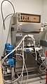
 Lab's FPLC.jpg 800 × 600; 85 kB
-
 Lac complementation.png 926 × 800; 74 kB
-
 Log2R.jpg 902 × 235; 30 kB
-
 MaPlot-edgeR.smear-wikipedia.png 648 × 432; 65 kB
-
 MeDIP (zh-cn).svg 1 150 × 1 000; 647 kB
-
 Medip-final-3.jpg 2 268 × 1 868; 349 kB
-
 MeDIP.svg 1 150 × 1 000; 648 kB
-
 Microarray-schema.gif 430 × 628; 30 kB
-
 Microarray2.gif 835 × 507; 133 kB
-
 MLPA (completa).png 748 × 906; 53 kB
-
 MST Technology.png 1 963 × 1 291; 338 kB
-
 Méthode de bertrand schema du dosage 2.jpg 236 × 305; 7 kB
-
 Nickel-resin regeneration.jpg 584 × 800; 130 kB
-
 Nihydrine réaction1.png 941 × 201; 14 kB
-
 Nihydrine réaction1.svg 1 250 × 275; 16 kB
-
 NMR sample.JPG 1 704 × 2 272; 651 kB
-
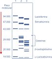 PAGE Lactoproteinas.jpg 1 125 × 1 258; 183 kB
-
 PB protein theory.svg 709 × 602; 27 kB
-
 PhOH-CHCl3 extraction.svg 268 × 179; 58 kB
-
 Photo 37.jpg 640 × 480; 82 kB
-
 Plotall mol.png 2 994 × 3 493; 372 kB
-
 Prinzip Sanger-Sequenzierung.png 536 × 448; 8 kB
-
 Protein-SDS interaction.png 1 679 × 544; 204 kB
-
 Prototype Terahertz Imager Promises Biochem Advances (5880464309).jpg 801 × 630; 566 kB
-
 PuceADN-schema FR.svg 430 × 628; 55 kB
-
 PuceADN-schema2 FR.svg 430 × 628; 55 kB
-
 Qiagen Mini Spin Column.svg 274 × 170; 48 kB
-
 RaPlot-jitter-wikipedia.png 648 × 432; 53 kB
-
 RaPlot-nouniques-wikipedia.png 648 × 432; 18 kB
-
 RaPlot-nouniques.jittered-wikipedia.png 648 × 432; 39 kB
-
 RaPlot-orange.uniques-wikipedia.png 648 × 432; 49 kB
-
 RaPlot-wikipedia.png 648 × 432; 23 kB
-
 Reactivo de Biuret.svg 598 × 243; 429 kB
-
 Reduced Representation Bisulfite Sequencing Protocol.jpg 10 048 × 4 097; 5,99 MB
-
 Resazurin resorufin dihydroresorufin.svg 1 567 × 290; 48 kB
-
 RIP-chip Overview.pdf 1 181 × 2 539; 47 kB
-
 RNASeqPics1.jpg 720 × 540; 46 kB
-
 RNASeqPics2.jpg 720 × 540; 33 kB
-
 Role of phosphate in urea-PAGE.svg 512 × 512; 44 kB
-
 Réduction dns.png 713 × 151; 11 kB
-
 Saltin in & Salting out.png 646 × 517; 105 kB
-
 Schematic of RNA chemical probing.png 700 × 1 400; 119 kB
-
 SDS-PAGE Electrophoresis.png 800 × 275; 40 kB
-
 SDS-PAGE overview.jpg 8 001 × 8 001; 8,53 MB
-
 SDS-PAGE sample.png 799 × 245; 35 kB
-
 SDS-PAGE.png 768 × 512; 260 kB
-
 Secuenciación con bisulfito.jpg 941 × 684; 87 kB
-
 Serial Dilution (to PCR).jpg 1 454 × 1 066; 104 kB
-
 Silac.gif 715 × 445; 12 kB
-
 SNAP-tag.svg 820 × 265; 100 kB
-
 SOFIA module.png 413 × 544; 30 kB
-
 SOFIA platform.jpg 2 592 × 1 936; 1,74 MB
-
 Southern-Blot-Autoradiogramm.jpg 196 × 304; 4 kB
-
 Ssap-vectors es.png 330 × 380; 11 kB
-
 Study-flow (zh-cn).svg 600 × 900; 330 kB
-
 Study-flow.jpg 984 × 1 508; 128 kB
-
 Study-flow.svg 600 × 900; 331 kB
-
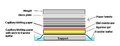
 Supports.png 475 × 254; 6 kB
-

-
 Western blot 2451.jpg 2 592 × 3 872; 765 kB
-
 Western Blot binding.png 796 × 284; 53 kB
-
 Western blot chemiluminescent detection.png 800 × 254; 33 kB
-
 Western blot transfer.png 799 × 283; 38 kB
-
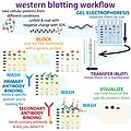 Western blot workflow.jpg 6 001 × 6 001; 7,01 MB
-
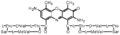
 CreatineSynthesis(en).png 2 892 × 898; 75 kB
-
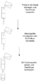
 Y2H copy.jpg 810 × 1 000; 161 kB


.jpg)
.jpg)
.jpg)
.jpg)


_-_DPLA_-_3f95e871f458451d2d00baa5a0d0d3fe.jpg)


.svg)


.png)


.jpg)


.jpg)


.svg)


.png)