Category:Cardiology
Salti al navigilo
Salti al serĉilo
Čeština: Kardiologie
· English: Cardiology
· Español: Cardiología
· Esperanto: Kardiologio
· Українська: Кардіологія
· medicina fako pri koro, arterioj, vejnoj, ktp. _es.svg) Diagrama del corazón humano. | |||||
| Alŝuti plurmedion | |||||
| Estas | |||||
|---|---|---|---|---|---|
| Subaro de |
| ||||
| Parto de | |||||
| Havas parton |
| ||||
| |||||

Subkategorioj
Ĉi tiu kategorio havas la 27 jenajn subkategoriojn, el 27 entute.
*
C
- Cerebrovascular circulation (6 D)
- Computational cardiology (2 D)
- Cardiology congresses (3 D)
D
H
L
M
- Cardiovascular models (93 D)
N
- Neocardiogenesis (2 D)
P
R
- Cardiac rehabilitation (1 D)
S
V
Dosieroj en kategorio “Cardiology”
La jenaj 179 dosieroj estas en ĉi tiu kategorio, el 179 entute.
-
De-Kardiologie.ogg 2,0 s; 20 KB
-
 1-s2.0-S2666602221000665-gr1 lrg.jpg 1 541 × 1 409; 206 KB
-
 An early cardiograph used by J.B.A. Chauveau. Photograph. Wellcome V0016529.jpg 3 232 × 2 444; 3,01 MB
-
 Anterior interventricular artery 2.png 2 550 × 3 350; 2,4 MB
-
 Anterior interventricular artery.png 2 550 × 3 350; 2,41 MB
-
 Anterior interventricular sulcus.png 2 550 × 3 350; 2,09 MB
-
 Anterior interventricular vein.png 2 550 × 3 350; 3,82 MB
-
 Anterior leaflet of left atrioventricular valve.png 2 550 × 3 350; 1,86 MB
-
 Anterior papillary muscle of right ventricle.png 2 550 × 3 350; 2,83 MB
-
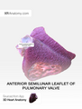 Anterior semilunar leaflet of pulmonary valve.png 2 550 × 3 350; 2,57 MB
-
 Anterior Sinus of pulmonary trunk.png 2 550 × 3 350; 3,59 MB
-
 Anterior surface of heart.png 2 550 × 3 350; 1,56 MB
-
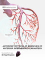 Anterior ventricular branches of anterior interventricular artery.png 2 550 × 3 350; 2,81 MB
-
 Anterior ventricular branches of right coronary artery.png 2 550 × 3 350; 3,14 MB
-
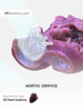 Aortic orifice.png 2 550 × 3 350; 4,11 MB
-
 Aortic Sinuses.png 2 550 × 3 350; 3,2 MB
-
 Aortic valve.png 2 550 × 3 350; 2,5 MB
-
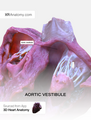 Aortic vestibule.png 2 550 × 3 350; 5,69 MB
-
 Apex of the heart.png 2 550 × 3 350; 1,23 MB
-
Athletic bradycardia wav.wav 20 s; 1,84 MB
-
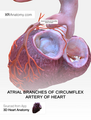 Atrial branches of circumflex artery of heart.png 2 550 × 3 350; 4,64 MB
-
 Atrial branches of right coronary artery.png 2 550 × 3 350; 2,73 MB
-
 Atrial flutter with 4-1 AV block.png 716 × 335; 194 KB
-
 Atrioventricular bundle.png 2 550 × 3 350; 2,34 MB
-
 Atrioventricular nodal branch of right coronary artery.png 2 550 × 3 350; 2,91 MB
-
 Atrioventricular node.png 2 550 × 3 350; 1,88 MB
-
 Atrioventricular septum.png 2 550 × 3 350; 2,28 MB
-
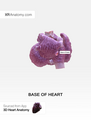 Base of heart.png 2 550 × 3 350; 1,76 MB
-
 Body of left atrium.png 2 550 × 3 350; 2,2 MB
-
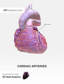 Cardiac arteries.png 2 550 × 3 350; 3,05 MB
-
 Cardiac sarcomere structure.png 2 903 × 1 417; 446 KB
-
 Cardiac septa.png 2 550 × 3 350; 1,72 MB
-
 Cardiac veins.png 2 550 × 3 350; 3,38 MB
-
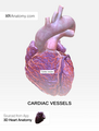 Cardiac vessels 2.png 2 550 × 3 350; 3,27 MB
-
 CardiacMuscle - longtitudinal.jpg 640 × 480; 188 KB
-
 Cardiographe.jpg 817 × 836; 105 KB
-
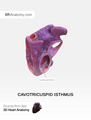 Cavotricuspid isthmus.png 2 550 × 3 350; 1,73 MB
-
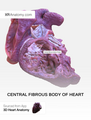 Central fibrous body of heart.png 2 550 × 3 350; 4,01 MB
-
 Chordae tendineae of left atrioventricular valve.png 2 550 × 3 350; 4,17 MB
-
 Chordae tendineae of right atrioventricular valve.png 2 550 × 3 350; 3,48 MB
-
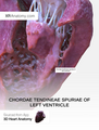 Chordae tendineae spuriae of left ventricle.png 2 550 × 3 350; 5,77 MB
-
 Circumflex artery of heart 2.png 2 550 × 3 350; 2,08 MB
-
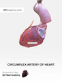
 Circumflex artery of heart.png 2 550 × 3 350; 3,91 MB
-
 Coarse apical trabeculations.png 2 550 × 3 350; 3,71 MB
-
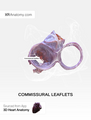 Commissural leaflets.png 2 550 × 3 350; 1,98 MB
-
 Commissures of semilunar leaflets of aortic valve.png 2 550 × 3 350; 2,44 MB
-
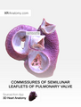 Commissures of semilunar leaflets of pulmonary valve.png 2 550 × 3 350; 2,92 MB
-
 Conal branch of anterior interventricular artery.png 2 550 × 3 350; 2,78 MB
-
 Conal branch of right coronary artery.png 2 550 × 3 350; 4,13 MB
-
 Conducting system of the heart.png 2 550 × 3 350; 1,68 MB
-
 Conus arteriosus.png 2 550 × 3 350; 2,93 MB
-
 Coronary sinus.png 2 550 × 3 350; 4,47 MB
-
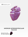
 Coronary sulcus.png 2 550 × 3 350; 2,45 MB
-
 Crista terminalis.png 2 550 × 3 350; 1,88 MB
-
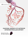 Diagonal branch of anterior interventricular artery.png 2 550 × 3 350; 2,33 MB
-
 Fibrous skeleton of heart.png 2 550 × 3 350; 1,92 MB
-
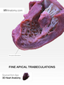 Fine apical trabeculations.png 2 550 × 3 350; 4,42 MB
-
 Fossa ovalis of right atrium.png 2 550 × 3 350; 2,86 MB
-
 Great cardiac vein.png 2 550 × 3 350; 3,93 MB
-
 Heart 1.tif 4 250 × 3 600; 912 KB
-
 Heart 2a.gif 4 250 × 3 600; 118 KB
-
 IFR-FFR Hybrid Approach.jpg 1 370 × 825; 160 KB
-
 Inferior border of heart.png 2 550 × 3 350; 1,75 MB
-
 Inferior interventricular artery.png 2 550 × 3 350; 2,78 MB
-
 Inferior interventricular sulcus.png 2 550 × 3 350; 1,96 MB
-
 Inferior leaflet of right atrioventricular valve.png 2 550 × 3 350; 2,15 MB
-
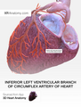 Inferior left ventricular branch of circumflex artery of heart.png 2 550 × 3 350; 4,88 MB
-
 Inferior papillary muscle of left ventricle.png 2 550 × 3 350; 6,26 MB
-
 Inferior papillary muscle of right ventricle.png 2 550 × 3 350; 3,45 MB
-
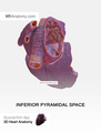 Inferior pyramidal space.png 2 550 × 3 350; 2,98 MB
-
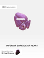 Inferior surface of heart.png 2 550 × 3 350; 1,52 MB
-
 Inferior vein of left ventricle.png 2 550 × 3 350; 3,44 MB
-
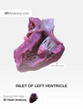 Inlet of left ventricle.png 2 550 × 3 350; 2,79 MB
-
 Inlet of right ventricle.png 2 550 × 3 350; 4,73 MB
-
 Interatrial septum.png 2 550 × 3 350; 2,63 MB
-
 Interleaflet triangles of aortic valve.png 2 550 × 3 350; 3,74 MB
-
 Interleaflet triangles of pulmonary trunk.png 2 550 × 3 350; 5,11 MB
-
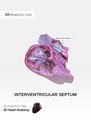 Interventricular septum.png 2 550 × 3 350; 2,23 MB
-
 Kardiomiocito sandara.jpg 563 × 568; 178 KB
-
 Left anterior branch of atrioventricular bundle.png 2 550 × 3 350; 2,34 MB
-
 Left atrioventricular (Mitral) valve.png 2 550 × 3 350; 2,1 MB
-
 Left atrioventricular orifice.png 2 550 × 3 350; 1,59 MB
-
 Left atrium.png 2 550 × 3 350; 2,39 MB
-
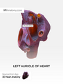 Left auricle of heart.png 2 550 × 3 350; 2,51 MB
-
 Left border of the heart.png 2 550 × 3 350; 1,46 MB
-
 Left bundle branch.png 2 550 × 3 350; 2,41 MB
-
 Left coronary aortic sinus.png 2 550 × 3 350; 4,13 MB
-
 Left coronary artery.png 2 550 × 3 350; 1,98 MB
-
 Left coronary leaflet.png 2 550 × 3 350; 2,18 MB
-
 Left fibrous ring.png 2 550 × 3 350; 3,71 MB
-
 Left fibrous trigone.png 2 550 × 3 350; 4,28 MB
-
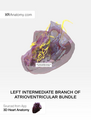 Left intermediate branch of atrioventricular bundle.png 2 550 × 3 350; 2,31 MB
-
 Left marginal branch of circumflex artery of heart.png 2 550 × 3 350; 3,45 MB
-
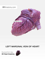 Left marginal vein of heart.png 2 550 × 3 350; 3,11 MB
-
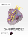 Left posterior branch of atrioventricular bundle.png 2 550 × 3 350; 2,63 MB
-
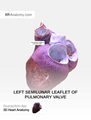 Left semilunar leaflet of pulmonary valve.png 2 550 × 3 350; 2,56 MB
-
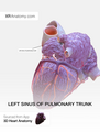 Left sinus of pulmonary trunk.png 2 550 × 3 350; 3,75 MB
-
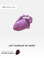 Left surface of heart.png 2 550 × 3 350; 1,58 MB
-
 Limbus fossae ovalis.png 2 550 × 3 350; 2,7 MB
-
 Localisation of the occlusion in STEMI.svg 716 × 354; 450 KB
-
 Lunules of semilunar leaflets of aortic valve.png 2 550 × 3 350; 2,49 MB
-
 Lunules of semilunar leaflets of pulmonary valve.png 2 550 × 3 350; 2,9 MB
-
 Membranous part of interventricular septum.png 2 550 × 3 350; 4,64 MB
-
 Middle cardiac vein.png 2 550 × 3 350; 4,48 MB
-
 Miokardas.jpg 360 × 240; 12 KB
-
 Multipulse Biowave Funktion.jpg 1 020 × 482; 44 KB
-
 Muscular part of interventricular septum.png 2 550 × 3 350; 3,93 MB
-
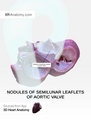 Nodules of semilunar leaflets of aortic valve.png 2 550 × 3 350; 2,58 MB
-
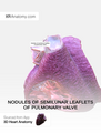 Nodules of semilunar leaflets of pulmonary valve.png 2 550 × 3 350; 3,31 MB
-
 Noncoronary aortic sinus.png 2 550 × 3 350; 3,31 MB
-
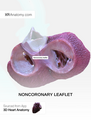 Noncoronary leaflet.png 2 550 × 3 350; 3,55 MB
-
 Oblique vein of left atrium.png 2 550 × 3 350; 3,38 MB
-
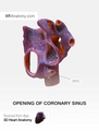 Opening of coronary sinus.png 2 550 × 3 350; 2,43 MB
-
 Opening of inferior vena cava.png 2 550 × 3 350; 2,61 MB
-
 Opening of pulmonary trunk.png 2 550 × 3 350; 2,4 MB
-
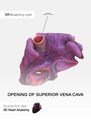 Opening of superior vena cava.png 2 550 × 3 350; 2,6 MB
-
 Openings of pulmonary veins.png 2 550 × 3 350; 2,48 MB
-

-
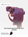 Pectinate muscles of left atrium.png 2 550 × 3 350; 2,85 MB
-
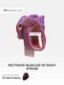 Pectinate muscles of right atrium.png 2 550 × 3 350; 2,13 MB
-
 Positron emission tomography.png 1 050 × 736; 531 KB
-
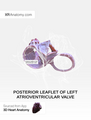 Posterior leaflet of left atrioventricular valve.png 2 550 × 3 350; 1,76 MB
-
 Pr Walinjom Muna.png 1 587 × 2 245; 1,5 MB
-
 Printvoorbeeld (CardioNetworks ECHOpedia).png 581 × 551; 28 KB
-
 Process of simultaneously recording ECG and SCG data.jpg 4 624 × 3 472; 4,87 MB
-
 PS61 reg.jpg 800 × 285; 102 KB
-
 PS61 Scheme.jpg 800 × 623; 52 KB
-
 Pulmonary arterial catheter.jpg 3 264 × 2 189; 1,79 MB
-
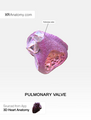 Pulmonary valve.png 2 550 × 3 350; 1,86 MB
-
 PV Loop loaded vs. unloaded.png 728 × 700; 743 KB
-
 Right atrioventricular (tricuspid) valve.png 2 550 × 3 350; 1,75 MB
-
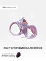 Right atrioventricular orifice.png 2 550 × 3 350; 1,88 MB
-
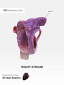 Right atrium.png 2 550 × 3 350; 1,87 MB
-
 Right auricle of heart.png 2 550 × 3 350; 2,32 MB
-
 Right border of heart.png 2 550 × 3 350; 1,43 MB
-
 Right bundle branch.png 2 550 × 3 350; 1,87 MB
-
 Right coronary aortic sinus.png 2 550 × 3 350; 4,26 MB
-
 Right coronary artery.png 2 550 × 3 350; 3,51 MB
-
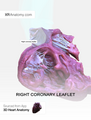 Right coronary leaflet.png 2 550 × 3 350; 4,07 MB
-
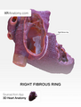 Right fibrous ring.png 2 550 × 3 350; 3,74 MB
-
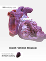 Right fibrous trigone.png 2 550 × 3 350; 3,81 MB
-
 Right inferolateral branch of right coronary artery.png 2 550 × 3 350; 2,55 MB
-
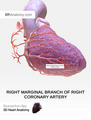 Right marginal branch of right coronary artery.png 2 550 × 3 350; 3,73 MB
-
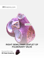 Right semilunar leaflet of pulmonary valve.png 2 550 × 3 350; 2,82 MB
-
 Right sinus of pulmonary trunk.png 2 550 × 3 350; 3,29 MB
-
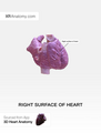 Right surface of heart.png 2 550 × 3 350; 1,5 MB
-
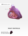 Right ventricle.png 2 550 × 3 350; 1,99 MB
-
 Root of Aorta.png 2 550 × 3 350; 4,55 MB
-
 Root of pulmonary trunk.png 2 550 × 3 350; 2,56 MB
-
 Septal branches of anterior interventricular artery.png 2 550 × 3 350; 3,16 MB
-
 Septal branches of inferior interventricular artery.png 2 550 × 3 350; 2,46 MB
-
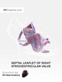 Septal leaflet of right atrioventricular valve.png 2 550 × 3 350; 1,91 MB
-
 Septal papillary muscle of right ventricle.png 2 550 × 3 350; 4,7 MB
-
 Septomarginal trabecula.png 2 550 × 3 350; 3,38 MB
-
 Sinuatrial (SA) node.png 2 550 × 3 350; 1,66 MB
-
 Sinuatrial nodal branch of right coronary artery.png 2 550 × 3 350; 3,83 MB
-
 Sinus of pulmonary veins.png 2 550 × 3 350; 3,14 MB
-
 Sinus of venae cavae.png 2 550 × 3 350; 2,57 MB
-
 Sinuses of pulmonary trunk.png 2 550 × 3 350; 3,65 MB
-
 Smooth part of left atrium.png 2 550 × 3 350; 3,18 MB
-
 Smooth part of right atrium.png 2 550 × 3 350; 2,12 MB
-
 Subendocardial branches.png 2 550 × 3 350; 1,9 MB
-
 Sulcus terminalis of the heart.png 2 550 × 3 350; 2,03 MB
-
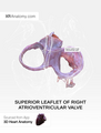 Superior leaflet of right atrioventricular valve.png 2 550 × 3 350; 2,19 MB
-
 Superior papillary muscle of left ventricle.png 2 550 × 3 350; 5,22 MB
-
 Supravalvular ridge of aorta.png 2 550 × 3 350; 4,83 MB
-
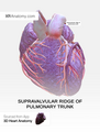 Supravalvular ridge of pulmonary trunk.png 2 550 × 3 350; 4,15 MB
-
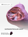 Supraventricular crest.png 2 550 × 3 350; 4,09 MB
-
 TAVRprocedure.jpg 183 × 275; 7 KB
-
 Tendon of inferior pyramidal space (Tendon of Todaro).png 2 550 × 3 350; 4,45 MB
-
 The journal of paediatrics, Jan-June 1945 Wellcome L0027013.jpg 1 658 × 1 116; 722 KB
-
 Trabeculae carneae of left ventricle.png 2 550 × 3 350; 3,1 MB
-
 Trabeculae carneae of right ventricle.png 2 550 × 3 350; 4,51 MB
-
 Trabecular part of left ventricle.png 2 550 × 3 350; 3,28 MB
-
 Trabecular part of right ventricle.png 2 550 × 3 350; 3,21 MB
-
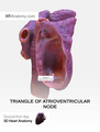 Triangle of atrioventricular node.png 2 550 × 3 350; 2,92 MB
-
 Unloaded PV Loop.png 545 × 498; 342 KB
-
 Эффективный циркуляционный объем31.jpg 1 268 × 1 938; 492 KB
-
 Սրտամկանի սարկոմերի կառուցվածքը.png 2 903 × 1 417; 437 KB


_valve.png)


_in_the_setting_of_an_accessory_AV_pathway.jpg)


.png)


_valve.png)


_node.png)


.png)


