Category:Cell nucleolus
Pereiti į navigaciją
Jump to search
largest structure in the nucleus of eukaryotic cells | |||||
| Įkelti mediją | |||||
| Tai yra |
| ||||
|---|---|---|---|---|---|
| Poklasis |
| ||||
| Yra dalis |
| ||||
| |||||
Daugialypės terpės rinkmenos kategorijoje „Cell nucleolus“
Rodomi 54 šios kategorijos rinkmenos (iš viso kategorijoje yra 54 rinkmenos).
-
-
-
-
 Allium-Differenzierung02-DM100x HF ba1.jpg 1 229 × 1 638; 1,73 MiB
-
 Allium-Differenzierung03-DM100x HF ba1.jpg 1 229 × 1 638; 1,42 MiB
-
 Allium-Differenzierung05-DM100x HF ba1.jpg 1 920 × 2 560; 1,62 MiB
-
 Apicomplexa structure.svg 450 × 207; 53 KiB
-
 Biological cell-2010-14-11.jpg 800 × 365; 86 KiB
-
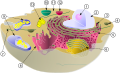 Biological cell.svg 1 466 × 891; 249 KiB
-
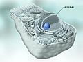
 Blausen 0212 CellNucleus ru.png 1 600 × 1 785; 2,15 MiB
-
 Blausen 0212 CellNucleus.png 1 600 × 1 785; 2,34 MiB
-
 Cafeteria roenbergensis FENCHEL and D J PATTERSON schematic drawing.svg 400 × 300; 13 KiB
-
 Cell nucleus-hu.png 1 600 × 1 785; 2,72 MiB
-
-
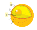 Diagram human cell nucleus numbered version.svg 462 × 378; 88 KiB
-
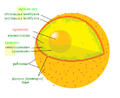 Diagram human cell nucleus serbian nucleolus.PNG 462 × 378; 46 KiB
-
 DL20240202 NCL-in-T98G.tif 9 977 × 3 780; 12,04 MiB
-
 DNA recycle hypothes.PNG 600 × 563; 259 KiB
-
 DnTRFc.jpg 462 × 259; 34 KiB
-
 Gangliocytic paraganglioma - 2 - intermed mag.jpg 2 848 × 4 272; 7,28 MiB
-
 Gangliocytic paraganglioma - high mag.jpg 2 848 × 4 272; 6,08 MiB
-
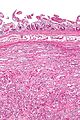 Gangliocytic paraganglioma - intermed mag.jpg 2 848 × 4 272; 5,82 MiB
-
 Gangliocytic paraganglioma - very high mag.jpg 2 848 × 4 272; 4,69 MiB
-
 HeLa-Tubulin-HSP60-Fibrillarin-DNA.jpg 2 968 × 2 976; 4,93 MiB
-
-
 Melanoma - cytology field stain.jpg 3 324 × 2 348; 3,05 MiB
-

-
Nucleolin-Inhibits-G4-Oligonucleotide-Unwinding-by-Werner-Helicase-pone.0035229.s004.ogv 20 s, 1 482 × 1 130; 5,32 MiB
-
 Nucleolus.jpg 640 × 480; 37 KiB
-
 NucleolusNCc.jpg 616 × 452; 120 KiB
-
 Nucleus Nucleolus and chromatin of animal cell.png 349 × 348; 245 KiB
-
 Nucleus&Nucleolus.gif 200 × 199; 42 KiB
-
 OSC Microbio 03 04 Nucleolus ku.png 883 × 384; 192 KiB
-
 OSC Microbio 03 04 Nucleolus.jpg 1 300 × 458; 426 KiB
-
 Ovocito pre-vitelogenico..png 574 × 330; 187 KiB
-

-
 P Cell.svg 640 × 640; 162 KiB
-
 Parasite160001-fig4 - Oogenesis in Crepidostomum metoecus (Digenea) TEM.png 2 392 × 1 178; 4,3 MiB
-
 Procryptobia glutinosa Cyst x49,500 TEM.jpg 2 095 × 2 345; 1,04 MiB
-
Reprogramming-of-Round-Spermatids-by-the-Germinal-Vesicle-Cytoplasm-in-Mice-pone.0078437.s001.ogv 38 s, 320 × 240; 295 KiB
-
Reprogramming-of-Round-Spermatids-by-the-Germinal-Vesicle-Cytoplasm-in-Mice-pone.0078437.s002.ogv 44 s, 320 × 240; 386 KiB
-
 Seminoma high mag.jpg 4 272 × 2 848; 4,83 MiB
-
 Seminoma intermed mag.jpg 4 272 × 2 848; 4,68 MiB
-
-
-
-
-
-
 Ultrastructure of choroid epithelium.jpg 1 200 × 1 000; 414 KiB
-
Widespread-Expression-of-BORISCTCFL-in-Normal-and-Cancer-Cells-pone.0022399.s008.ogv 20 s, 1 408 × 788; 1,35 MiB
-
Widespread-Expression-of-BORISCTCFL-in-Normal-and-Cancer-Cells-pone.0022399.s009.ogv 20 s, 1 408 × 788; 4,88 MiB
-
Widespread-Expression-of-BORISCTCFL-in-Normal-and-Cancer-Cells-pone.0022399.s010.ogv 20 s, 1 544 × 788; 909 KiB
-
Widespread-Expression-of-BORISCTCFL-in-Normal-and-Cancer-Cells-pone.0022399.s011.ogv 20 s, 1 568 × 788; 5,61 MiB
-
 خانە و بەشەکانی.jpg 673 × 513; 67 KiB


_TEM.png)


_(white)_in_the_cytoplasm_(green)_of_clusters_of_conifer_cells_one_hour_after_mechanical_agitation.jpg)

