Category:Cochlea
Vai alla navigazione
Vai alla ricerca
componente dell'orecchio interno | |||||
| Carica un file multimediale | |||||
| Istanza di |
| ||||
|---|---|---|---|---|---|
| Sottoclasse di |
| ||||
| Parte di |
| ||||
| Uso | |||||
| Consiste di |
| ||||
| |||||
Sottocategorie
Questa categoria contiene le 4 sottocategorie indicate di seguito, su un totale di 4.
File nella categoria "Cochlea"
Questa categoria contiene 45 file, indicati di seguito, su un totale di 45.
-
1406 Cochlea ku.jpg 913 × 548; 103 KB
-
A spatial gradient of S100+SOX9+ Peripheral Glial Cells in the W12 human fetal cochlea.png 2 387 × 1 352; 1,83 MB
-
Abgerollte-schnecke.png 3 240 × 1 424; 245 KB
-
Basilair membraan bovenaanzicht.jpg 736 × 750; 141 KB
-
Basilar membrane uncoiled detail.jpg 2 152 × 1 896; 598 KB
-
Capturing PGCs in the human cochlea.png 1 993 × 4 413; 1,28 MB
-
Cel ciliadas Coclea.png 1 661 × 1 873; 1,37 MB
-
Ch14 cochlea.png 542 × 375; 67 KB
-
Chochlear amplifier (using Prestin motor proteins).jpg 2 200 × 1 588; 192 KB
-
Cochlea inner ear.svg 409 × 423; 108 KB
-
Cochlea wave animated.gif 200 × 143; 11 KB
-
Coclea 3D.png 981 × 935; 564 KB
-
Cupula cs.png 512 × 480; 47 KB
-
Cóclea desespiralada 1.png 1 742 × 274; 35 KB
-
Cóclea desespiralada 2.png 1 742 × 397; 47 KB
-
Cóclea desespiralada 3.png 1 742 × 506; 54 KB
-
Cóclea desespiralada 4.png 1 742 × 451; 59 KB
-
Cóclea desespiralada 5.png 1 548 × 416; 198 KB
-
Ductus cochlearis schema 2-esp.jpg 981 × 851; 150 KB
-
Ductus cochlearis schema 2.jpg 981 × 851; 145 KB
-
Ductus cochlearis schema esp.jpg 981 × 851; 190 KB
-
Ductus cochlearis schema.jpg 981 × 851; 151 KB
-
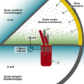
Elektrische Potentiale im Innenohr.png 2 300 × 2 300; 497 KB
-
Estructura coclear.jpg 911 × 476; 89 KB
-
Green fluorescent protein image of the mouse cochlea.jpg 400 × 400; 29 KB
-
Helicotrema 2.png 1 200 × 527; 130 KB
-
Helicotrema.jpg 3 240 × 1 424; 447 KB
-
Immature Schwann cells along the peripheral processes express NGFR.png 1 248 × 1 703; 2,72 MB
-
Janela redonda.webm 10 s, 1 280 × 720; 268 KB
-
Journey of Sound to the Brain.ogv 2 min 27 s, 1 280 × 720; 40,92 MB
-
NGFR expression in the cochlear duct epithelium.png 1 949 × 1 807; 2,66 MB
-
Noise transmission paths through cochlea.jpg 581 × 365; 107 KB
-
Peripheral Glial Cells expressed SOX10, SOX9 and S100B in the W10.4 human fetal cochlea.png 1 319 × 1 663; 1,93 MB
-
Peripheral Glial Cells expressed SOX10, SOX9 and S100B in the W9 human fetal cochlea.png 1 808 × 1 577; 4,5 MB
-
Schematic uncoiled cochlea.png 1 993 × 347; 70 KB
-
Snail cochlea.jpg 1 080 × 1 287; 479 KB
-
Stria vascularis1.jpg 1 057 × 1 078; 164 KB
-
Terminal differentiation of Schwann cells in the cochlear nerve.png 1 253 × 2 505; 4,74 MB
-
Two Views of Cochlear Mechanics.jpg 2 176 × 2 688; 401 KB
-
Udito.webm 2 min 27 s, 1 280 × 720; 31,88 MB
-
Uncoiled cochlea schematic.jpg 926 × 297; 65 KB
-
Vibrations of basilar membrane.jpg 1 000 × 1 316; 297 KB
-
Wie funktioniert unser Gleichgewichtssinn?.webm 1 min 16 s, 1 920 × 1 080; 99,55 MB
-
قوقعة حلزون 2.jpg 3 000 × 4 000; 3,08 MB


