Category:Computed tomography
Pereiti į navigaciją
Jump to search

medical imaging procedure using X-rays to produce cross-sectional images  | |||||
| Įkelti mediją | |||||
| Tai yra |
| ||||
|---|---|---|---|---|---|
| Poklasis | |||||
| |||||
Subkategorijos
Rodomos 24 subkategorijos (iš viso yra 24 subkategorijos).
3
4
- 4DCT (2 F)
A
- Artifacts in computed tomography (11 F)
C
- Cinematic rendering (4 F)
- Computed tomography schematics (54 F)
D
F
- Filtered backprojection (28 F)
I
- Iterative reconstruction (18 F)
M
- Mobile stroke unit (4 F)
O
- Oral computed tomography (11 F)
P
S
- Shepp-Logan phantom (8 F)
- Sinogram (15 F)
X
Daugialypės terpės rinkmenos kategorijoje „Computed tomography“
Rodomi 66 šios kategorijos rinkmenos (iš viso kategorijoje yra 66 rinkmenos).
-
De-Computertomographie.ogg 2,1 s; 20 KiB
-
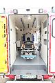 2016.05.04 MSU, Mobile Stroke Unit interior.jpg 2 304 × 3 456; 2,42 MiB
-

-
 Bga.jpg 227 × 161; 9 KiB
-
 Blausen 0206 CATScan 02.png 768 × 1 024; 2,25 MiB
-
 Casting porosity.jpg 160 × 115; 7 KiB
-
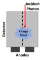 Charge Sharing.png 700 × 962; 32 KiB
-

-
 Cone Beam CT Scanner.png 445 × 211; 166 KiB
-
 CT Halo sign around a right lower lobe pulmonary nodule.png 512 × 204; 124 KiB
-
 CT Head Topogram.jpg 512 × 512; 178 KiB
-
 CT image guided injection pudendal nerve.png 2 220 × 1 498; 1,03 MiB
-
 CT of human thorax showing current paths for EIT corrected.jpg 613 × 613; 76 KiB
-
 CT presentation as thin slice, projection and volume rendering.jpg 1 190 × 1 035; 302 KiB
-
 CT presentation as thin slice, projection and volume rendering.svg 1 115 × 970; 2,38 MiB
-
 CT Salt Mill.jpg 1 725 × 7 175; 3,39 MiB
-
 CT Scan Liver-Kidneys.jpg 5 017 × 3 345; 1,54 MiB
-

-
 Diagnostika-raka 4.jpg 638 × 443; 34 KiB
-
 Diffusivitaet FA versus RA.jpg 400 × 300; 30 KiB
-
 Diffusivitaet Fraktionale Anisotropie.jpg 400 × 300; 33 KiB
-
 Diffusivitaet Relative Anisotropie.jpg 400 × 300; 32 KiB
-
 Diffusivitaet VV.jpg 400 × 300; 32 KiB
-
 Fairfield CT Precincts 2015-2021.svg 1 200 × 800; 10 KiB
-
 First Generation CT Scan.svg 602 × 602; 17 KiB
-
 Fourth Generation CT Scan.svg 602 × 602; 319 KiB
-
 Generation of STL - 3.jpg 740 × 411; 26 KiB
-
 Generation of STL.jpg 160 × 89; 3 KiB
-
 Ilustration showing FBP, Hybrid IR and Model-based IR.svg 617 × 458; 623 KiB
-
 Medicoplacas.JPG 3 216 × 2 136; 4,5 MiB
-
 Micro-CT braided polymer rope 3D 11.jpg 2 139 × 1 451; 2,5 MiB
-
 Mobile-stroke-unit-exterior.jpg 4 000 × 2 611; 1,18 MiB
-
 MobiScan House.jpg 1 920 × 1 080; 945 KiB
-
 Nameplate of a CT.jpg 4 608 × 2 592; 1,75 MiB
-
 NCCT Abdomen.jpg 390 × 318; 37 KiB
-
 Nigoi-CT-Q08937-thumb.jpg 900 × 600; 53 KiB
-
 NM19 102.gif 442 × 271; 6 KiB
-
 NM19 106.gif 460 × 525; 7 KiB
-
 NM19 107a.gif 340 × 272; 2 KiB
-
 Ors-visual-vessel-analysis.png 1 090 × 721; 709 KiB
-
 Ovarian Squamous Carcinoma Tumor.jpg 1 969 × 1 992; 316 KiB
-
 Pharmacological Capsule Diclofenac μCT.jpg 1 761 × 10 150; 5,88 MiB
-
 Pulse Pileup.png 654 × 752; 32 KiB
-
 Radon transform example.jpg 1 257 × 612; 52 KiB
-
 Reconstructions in dual-energy and photon-counting computed tomography.svg 627 × 383; 565 KiB
-
 Robert S. Ledley's Computer Aided Tomography Patent - Figure 3.png 3 408 × 2 320; 70 KiB
-
 Robert S. Ledley's Computer Aided Tomography Patent - Figures 1 and 2.png 2 320 × 3 408; 97 KiB
-
 Robert S. Ledley's Computer Aided Tomography Patent - Figures 4 and 5.png 3 408 × 2 320; 71 KiB
-
 Robert S. Ledley's Computer Aided Tomography Patent - Figures 6 to 10.png 3 408 × 2 320; 76 KiB
-
 Robert S. Ledley's Computer Aided Tomography Patent - Figures IIA, IIB and IIC.png 2 320 × 3 408; 40 KiB
-
 Searching for a cause (14061196032).jpg 1 024 × 679; 216 KiB
-
 Second Generation CT Scan.svg 602 × 602; 389 KiB
-
 Sinuses and Sinusitis (5937085231).jpg 559 × 695; 288 KiB
-
 Third generation CT.svg 602 × 602; 340 KiB
-

-
 Tibia-CT-Biomechanics-part1.png 990 × 814; 362 KiB
-
 Tibia-CT-Biomechanics-part2.png 1 119 × 701; 392 KiB
-
Tomografía axial computarizada.wav 51 s; 8,56 MiB
-
 TomographyPrinciple Illustration.png 794 × 1 123; 91 KiB
-
 TomoScope XS Geräte.png 1 181 × 788; 4,16 MiB
-
 TORNAI-SpectrumOfMedicalImaging.jpg 720 × 504; 117 KiB
-
 Unknownvalues.PNG 273 × 323; 7 KiB
-
 VX1 DICOM.jpg 1 024 × 681; 74 KiB
-
 Xray examination.jpg 2 322 × 4 128; 2,5 MiB
-
 ZEISS Crossbeam (9662745943).jpg 8 858 × 4 992; 3,22 MiB
-
 Átvilágított tárgy és intenzitásgörbe.PNG 313 × 196; 13 KiB


_versus_X-ray_microtomography_(X-ray_%CE%BCCT)_of_a_Lego_minifigure_-_1-s2.0-S2949673X22000018-gr4_lrg.jpg)


.jpg)

.jpg)


.jpg)
