Category:Diseases and disorders of the eye and adnexa
Pereiti į navigaciją
Jump to search
Radiologija: Ultragarsas · Kompiuterinė tomografija · Magnetic resonance | Anatomical pathology: Gross pathology · Histopathology | Other: Akies dugnas · Optical coherence tomography · Epidemiologija (žemėlapis → Pasaulis) | File format: SVG · Video | 

Categorization according to International Statistical Classification of Diseases and Related Health Problems, 10th Revision.
[keisti]health condition negatively affecting the eye | |||||
| Įkelti mediją | |||||
| Tai yra |
| ||||
|---|---|---|---|---|---|
| Poklasis |
| ||||
| |||||
Subkategorijos
Rodomos 55 subkategorijos (iš viso yra 55 subkategorijos).
*
-
A
- Adie syndrome (1 F)
B
- Bitot's spots (2 F)
- Buphthalmos (2 F)
C
- Corectopia (1 F)
- Eye cysts (2 F)
D
F
G
- Globe rupture (3 F)
- Eye gumma (2 F)
H
I
K
- Keratic precipitate (2 F)
L
- Leukocoria (5 F)
N
O
P
S
- Scleromalacia perforans (1 F)
V
W
- Waardenburg syndrome (18 F)
Puslapiai kategorijoje „Diseases and disorders of the eye and adnexa“
Šioje kategorijoje yra vienas puslapis.
Daugialypės terpės rinkmenos kategorijoje „Diseases and disorders of the eye and adnexa“
Rodomi 39 šios kategorijos rinkmenos (iš viso kategorijoje yra 39 rinkmenos).
-
 120823-F-CF823-217 (7938218002).jpg 4 256 × 2 832; 6,01 MiB
-
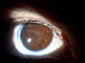
 2012-09-22 eye with disease.jpg 2 768 × 2 079; 958 KiB
-
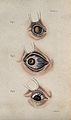 A sheet of three diagrams showing inflamed bloodshot eye def Wellcome V0015923EL.jpg 1 266 × 2 136; 1,53 MiB
-
 A sheet of three diagrams showing inflamed bloodshot eye def Wellcome V0015923ER.jpg 1 233 × 2 086; 1,32 MiB
-
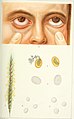
 A sheet of three diagrams showing inflamed eye defects. Colo Wellcome V0015922EL.jpg 1 272 × 2 082; 1,71 MiB
-
 A sheet of three diagrams showing inflamed eye defects. Colo Wellcome V0015922ER.jpg 1 251 × 2 088; 1,49 MiB
-
 Acoria pupilar.jpg 2 048 × 1 536; 1,2 MiB
-
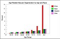 AMD by race and age from National Eye Institute data.jpg 1 882 × 1 216; 289 KiB
-

-
 Bilateral222.jpg 666 × 247; 94 KiB
-
 Eye defect teaching model, on wood stand, probably English, Wellcome L0057876.jpg 4 709 × 2 232; 776 KiB
-
 Eye defect teaching model, on wood stand, probably English, Wellcome L0057877.jpg 4 256 × 2 832; 782 KiB
-
 Eye defect teaching model, on wood stand, probably English, Wellcome L0057878.jpg 4 256 × 2 832; 1,33 MiB
-
 Eye discharge.jpg 1 690 × 1 044; 321 KiB
-

-

-
-
 Irvine-Gass syndrome .png 1 920 × 1 329; 538 KiB
-
 KITLV A434 - Patiënte met een oogziekte in Nederlands-Indië, KITLV 35584.tiff 792 × 757; 1,72 MiB
-
 Macro Globe oculaire - Mélanome 55-o.apatho-52a-oeil.jpg 639 × 1 499; 500 KiB
-
 Macro Globe oculaire - Mélanome 55-o.apatho-52p-oeil.jpg 735 × 540; 195 KiB
-
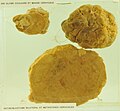 Macro Globe oculaire - Rétinoblastome 55-o.apatho-242a-oeil.jpg 1 676 × 1 547; 1,44 MiB
-
 Macro Globe oculaire - Rétinoblastome 55-o.apatho-242p-oeil.jpg 1 563 × 1 497; 1,33 MiB
-
 Man with large, malignant growths protruding from both orbits Wellcome L0061863.jpg 4 404 × 5 196; 3,84 MiB
-
 Melanosis of the eyeball and orbit Wellcome L0061862.jpg 6 160 × 4 048; 4,4 MiB
-
 Membranous conjunctivitis.jpg 2 023 × 1 125; 570 KiB
-
 Nikki Wordsmith Odd Eyes.jpg 828 × 570; 360 KiB
-
 Olho com conjuntivite bacteriana.jpg 711 × 1 073; 441 KiB
-
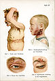
 Pediatrics. (1902) (14577803960).jpg 2 218 × 3 472; 856 KiB
-
 PieIXretDiab.jpg 2 322 × 2 224; 923 KiB
-
 Pupil of an eye occluded by lymph Wellcome L0061851.jpg 6 012 × 3 788; 2,47 MiB
-
 Reke Salat.png 181 × 216; 50 KiB
-
 Retrobulbarbleed.jpg 595 × 552; 102 KiB
-
 Siebert 28.jpg 1 945 × 2 866; 1,13 MiB
-
 Tape removal.jpg 818 × 615; 110 KiB
-
 Vision alterada por SOM diurno.jpg 1 289 × 780; 641 KiB
-
 Vision alterada por SOM nocturno.jpg 1 063 × 672; 395 KiB
-
 Yamai no Soshi - Eye Disease (part 1).jpeg 2 420 × 2 368; 3,19 MiB
-
 Ülekoormuse põhjustatud paistetus.jpg 2 859 × 1 906; 3,3 MiB
.jpg)


_(14596082218).jpg)


.png)
_Hrsg._von_W._Kolle_und_A._von_Wassermann_(1912-13)_(16662993341).jpg)


_(14577803960).jpg)


.jpeg)
